FIDEX+ European Premiere |WSAVA Warsaw |13–15 October 2026 |Booth 69 Book a Meeting
Animage
EN Request a Call
{{ panelLabel }}
{{ panelSub }}
{{ panelFoot }}
Home / Software & Workflow
Connected veterinary imaging

From patient setup to clinical review-one connected workflow.

FIDEX brings image acquisition, reconstruction, visualisation, study management and sharing into one veterinary-focused working environment.

By connecting multiple imaging modalities with an integrated software workflow, FIDEX helps clinical teams move efficiently from examination setup to completed study.

{{ swStep }}
{{ swLabel }}
The interface

One application. Four tabs. Every modality.

Database, DR, CT and Fluoroscopy sit as four tabs in the same window, with acquisition parameters on one side and the live study on the other. These are the actual FIDEX screens, not mock-ups - click any of them to read the detail at full size.
DR acquisition - FIDEX interface DR · Acquisition View full size ↗
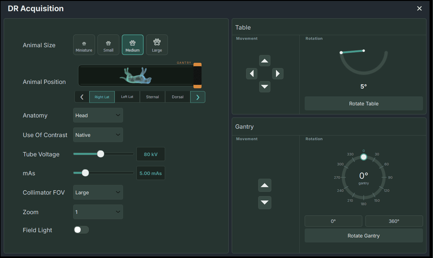
DR acquisition
Animal size and position, anatomy, contrast, collimator and field light - with table and gantry movement alongside.
This study: 80 kV · 5.00 mAs · large collimator FOV
CT acquisition - FIDEX interface CT · Acquisition View full size ↗
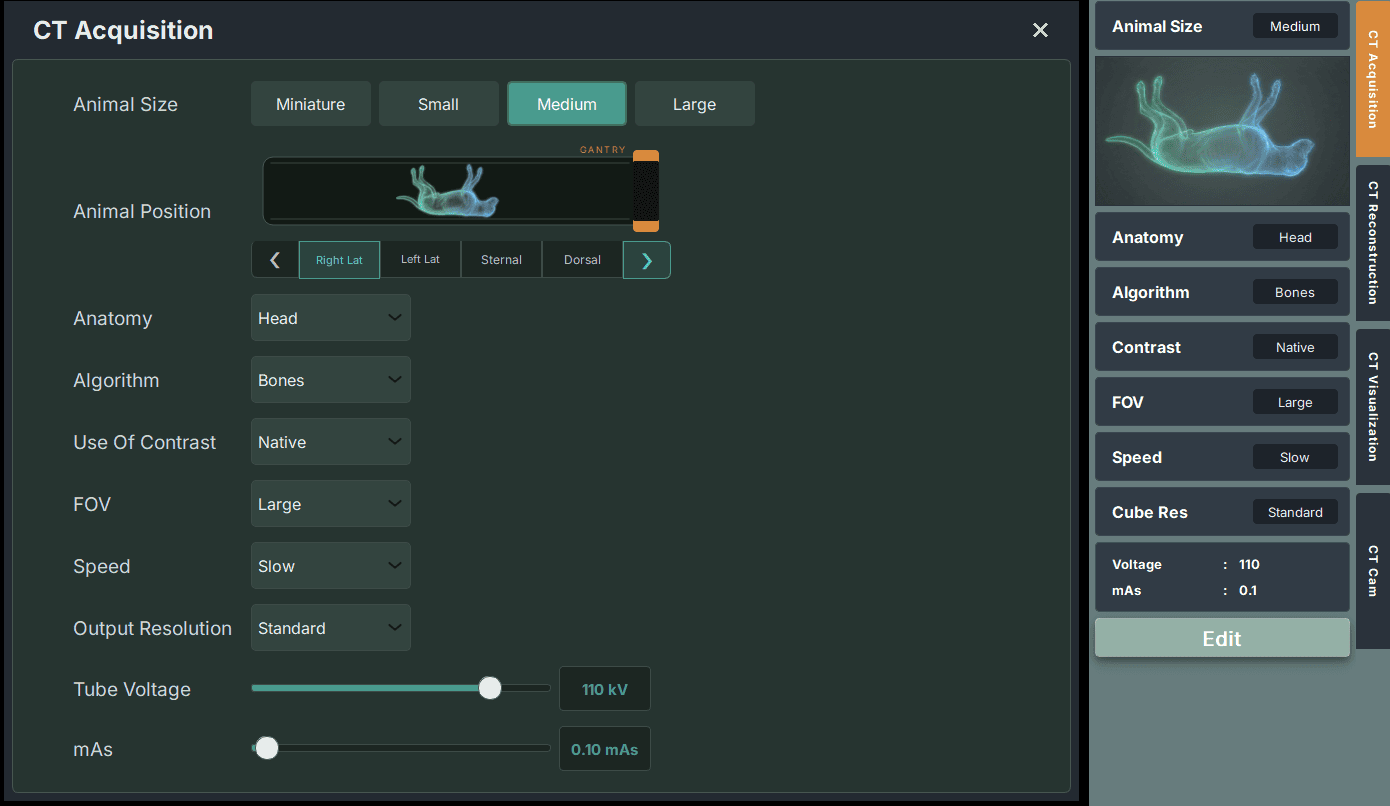
CT acquisition
Protocol-led. Anatomy, reconstruction algorithm, field of view, speed and output resolution are all set before the scan starts.
This study: 110 kV · 0.10 mAs · bone algorithm
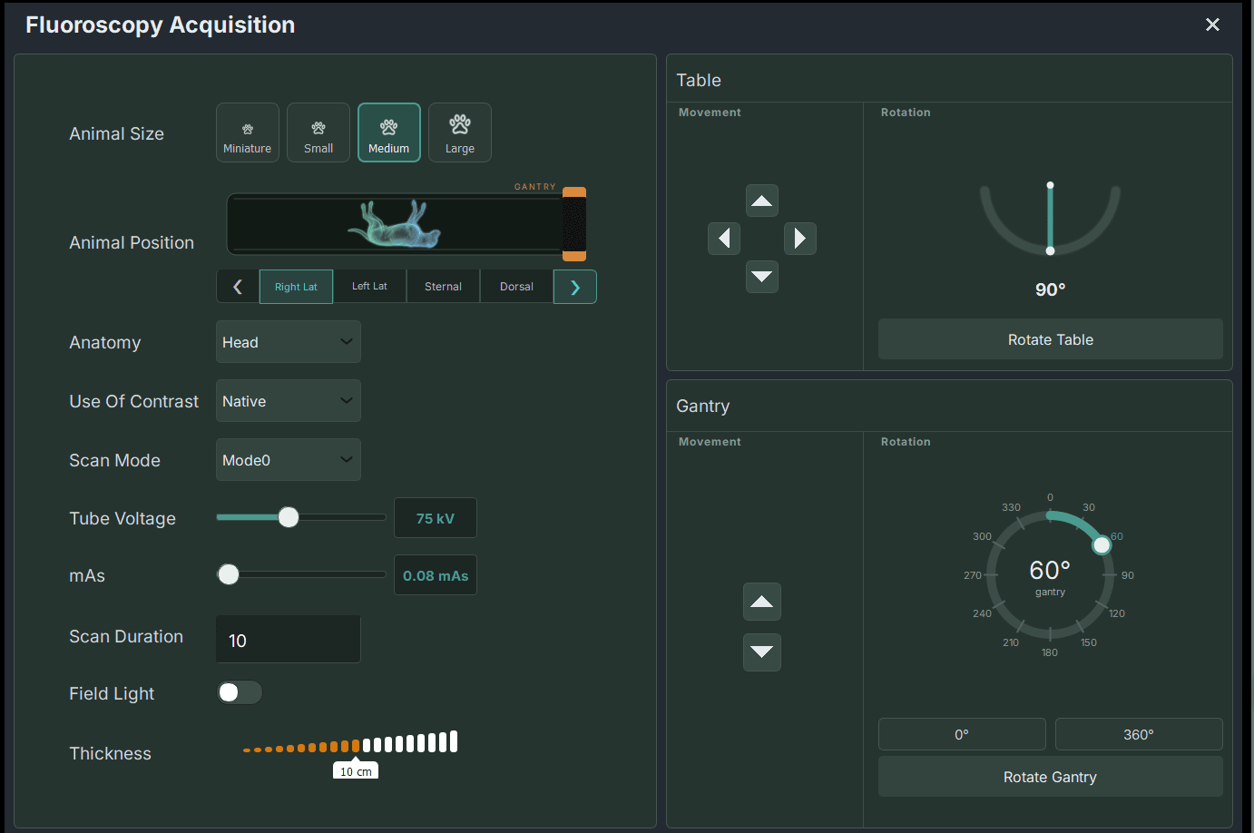
Fluoroscopy acquisition - FIDEX interface FL · Acquisition View full size ↗
Fluoroscopy acquisition
Scan mode, duration and thickness, with independent table and gantry rotation for the projection you need.
This study: 75 kV · 0.08 mAs · 10 s · 100 frames · gantry 60°
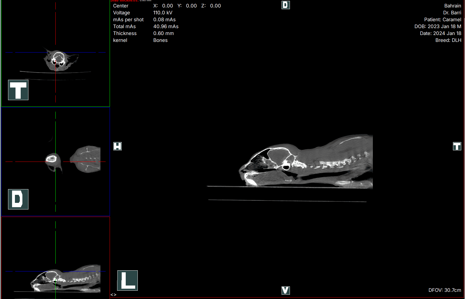
CT reconstruction and MPR - FIDEX interface CT · Review View full size ↗
CT reconstruction and MPR
Transverse, dorsal and sagittal planes together with the primary view. MIP, MPR, slab and slice modes, measurement, annotation and saveable presets.
This study: feline head · 0.60 mm slab · 40.96 total mAs · DFOV 30.7 cm
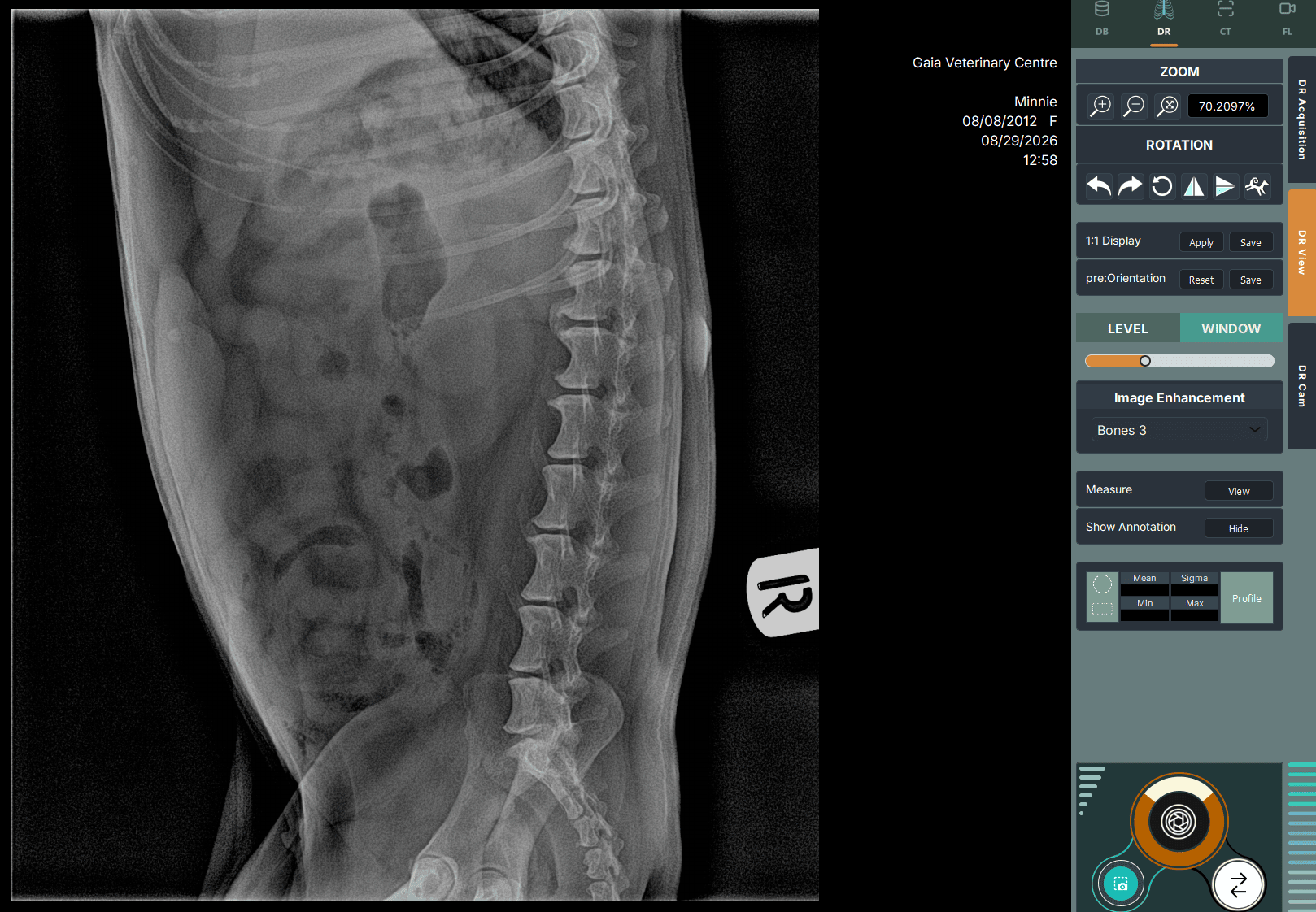
DR review - FIDEX interface DR · Review View full size ↗
DR review
Zoom, rotation, level and window, image-enhancement presets, measurement and annotation - all beside the image.
This study: canine abdomen · 75 kV · 4.00 mAs · Bones 3
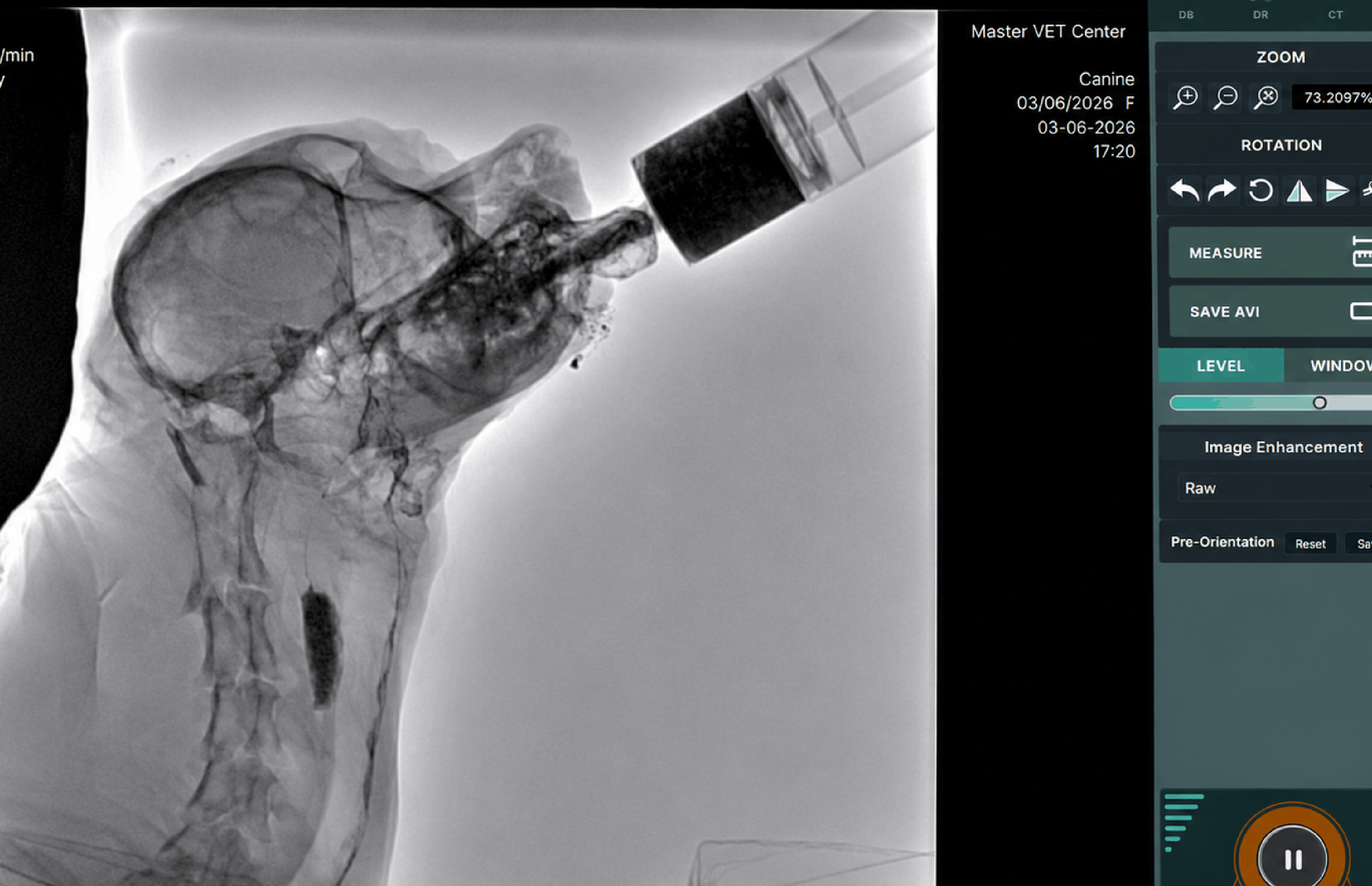
Fluoroscopy review - FIDEX interface FL · Review View full size ↗
Fluoroscopy review
Real-time playback with frame stepping, live dose readout and AVI export for the clinical record.
This study: canine contrast study · 65 kV · AKR 4.32 mGy/min · CumAX 12.87 mGy
Patient database - FIDEX interface DB · Patients View full size ↗
Patient database
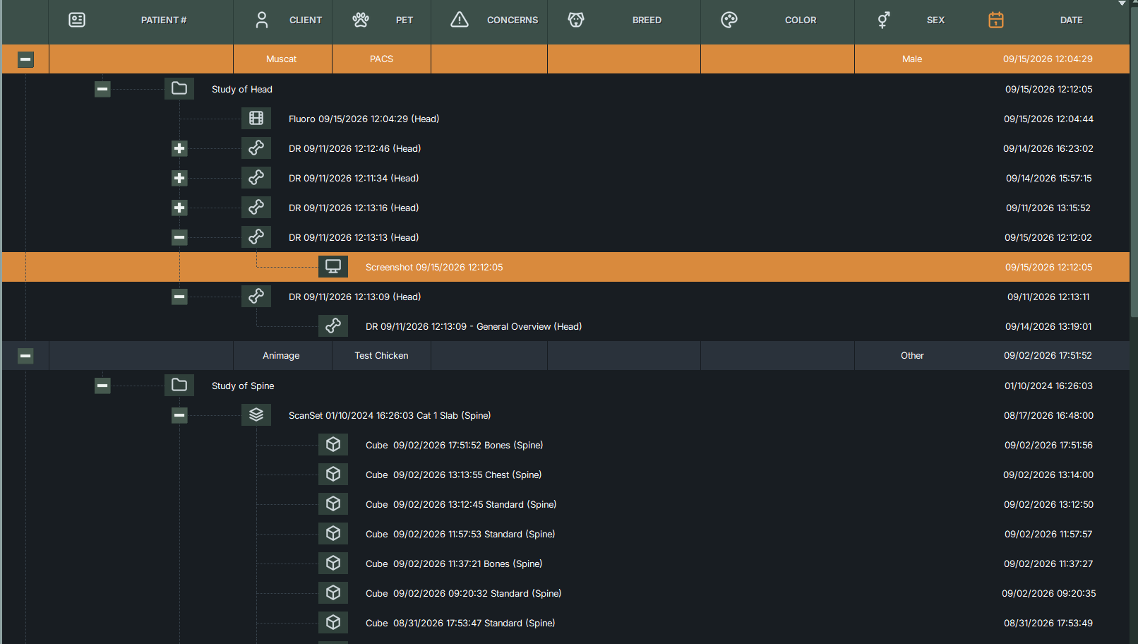
Patient, client, pet, concerns, breed, colour and sex - with every study, ScanSet and reconstruction nested beneath.
DICOM send queue - FIDEX interface DB · DICOM View full size ↗
DICOM send queue
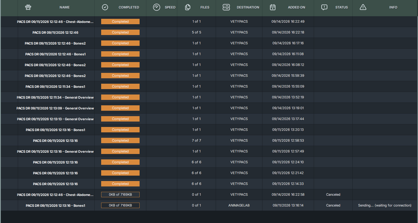
Studies pushed to any DICOM destination, with per-job progress, file counts and status.
Destinations shown: VETYPACS, ANIMAGELAB

Screens captured from a FIDEX+ system. The parameter values above are those of the example study, not the limits of the system - confirmed ranges appear on each product page.

Workflow overview

Designed around the imaging journey.

The exact workflow varies by product, configuration and examination, but a typical FIDEX study may follow seven connected stages.

{{ activeStage.no }}
Stage {{ activeStage.no }} of 7

{{ activeStage.name }}

{{ activeStage.desc }}

Veterinary-focused operation

Technology that supports the examination-not distracts from it.

FIDEX software is intended to help veterinary teams manage complex imaging tasks through a more structured operating environment.

Depending on the selected product, capabilities may include:

{{ it.n }}
{{ it.label }}
Multi-modality workflow

Move between imaging capabilities within one platform.

FIDEX combines CT, Digital Radiography and Fluoroscopy within one imaging environment. This can allow the veterinary team to perform complementary examinations without transferring the patient between separate systems.

{{ m.n }}
{{ m.tag }}

{{ m.desc }}

Not every case requires every modality. The veterinary team determines the appropriate examination according to the patient and clinical question.

CT reconstruction

Review anatomy from multiple perspectives.

CT acquisition produces volumetric data that can be reconstructed and reviewed in different formats. Depending on the FIDEX product and software configuration, available tools may include:

{{ r.n }}

{{ r.name }}

{{ r.desc }}

View Clinical Imaging Examples →
Digital Radiography workflow

Efficient imaging for everyday clinical use.

Digital Radiography is integrated into the FIDEX working environment, supporting routine imaging, patient positioning and clinical follow-up.

Available functionality may include
{{ it.n }}
{{ it.label }}
Fluoroscopy and DSA

Real-time imaging when movement matters.

Fluoroscopy supports dynamic examinations and selected image-guided procedures. Depending on the product configuration, functionality may include:

{{ it.n }}
{{ it.label }}

Clinical use should follow appropriate veterinary protocols, radiation-safety procedures and operator training.

ABC™ Automated Breathing Control

Supporting respiratory-motion management.

01
The challenge
Respiratory motion can affect image quality during veterinary CT examinations, particularly in the thorax and abdomen.
02
How ABC™ helps
ABC™ Automated Breathing Control is designed to coordinate image acquisition with compatible veterinary ventilation workflows during selected studies.
03
Who stays in control
The feature is intended to help the veterinary team manage breathing-related motion while maintaining control of the examination.
Learn About ABC™ →
Contrast workflow

Support for selected contrast-enhanced examinations.

Selected FIDEX configurations may support contrast-study workflows through CT, Fluoroscopy, DSA and Auto Contrast Tracking functionality.

Potential uses may include
{{ it.n }}
{{ it.label }}

Auto Contrast Tracking and Cine-CT availability should be identified by product rather than presented as universal FIDEX functionality.

DICOM and PACS connectivity

Connect imaging with the wider clinical environment.

FIDEX systems are intended to support DICOM-based imaging workflows. Depending on the product, software version and local infrastructure, integration may include:

{{ g.n }}
{{ g.title }}

{{ g.intro }}

{{ it }}
Discuss Integration Requirements Request Technical Documentation
FIDEX+ and the future workflow

Building the next generation of assisted imaging.

FIDEX+ is intended to evolve the FIDEX workflow through new acquisition, positioning, control and software capabilities.

Early 2027
FIDEX+ table-side controls
Table, U-arm and mode at the patient's side
Human-led
AI-supported features should assist the clinical team without replacing veterinary judgement or radiological interpretation.
Explore FIDEX+ →
Proposed functionality may include
{{ it.n }}
{{ it.label }}
Training and implementation

Help your team make the most of the workflow.

Depending on product and region, implementation support may include:

FIDEX in a standard clinical room
2
Before installation
3
At go-live
4
Ongoing
Explore Training and Support →
{{ it.n }}
{{ it.label }}
{{ it.phase }}
FAQ

Frequently Asked Questions

{{ f.q }}

{{ f.a }}

See the complete FIDEX workflow.

A demonstration can show how patient setup, modality selection, image acquisition, reconstruction, review and sharing work together.

Request a Workflow Demo Discuss Integration Requirements