A crisp-looking scan isn’t necessarily a diagnostic one. Veterinary CBCT image quality depends on the full imaging process, including patient motion, anatomy, positioning, scan settings and reconstruction. Bone edges may appear sharp while soft-tissue contrast remains limited. Scatter, beam hardening or metal can also obscure detail that matters to the clinical question.
That distinction can be difficult to judge when technical terms are unfamiliar and a visually clear scan seems reassuring. The key is to assess what the image can show for the specific diagnostic task, not clarity alone. This article explains the factors that shape image quality, how to recognize common artefacts and interpretive limitations, and how to assess a scan consistently in clinical context. It covers spatial resolution, contrast and noise, motion, anatomy and acquisition choices, then brings them together in a practical framework for deciding whether an image is fit for its intended purpose.
Key Takeaways
- Judge veterinary CBCT image quality by how well the scan supports the clinical question, not by apparent sharpness alone.
- Consider acquisition and reconstruction together, including whether the field of view includes the region and structures that need assessment.
- Recognize that artefacts can obscure, distort or imitate anatomy, and that their causes and effects vary.
- Use a consistent review sequence: define the question, confirm the region imaged, then assess image characteristics and limitations.
- FIDEX+ combines veterinary CBCT, digital radiography and fluoroscopy in one imaging platform. This combination does not imply a specific image-quality outcome.
What Does Veterinary CBCT Image Quality Actually Mean?
Image quality isn’t a single measure of how crisp a scan looks. It describes how well the image supports assessment of a specific clinical question. A volume can show sharply defined bone yet provide limited information about soft tissues. A scan that looks clear at first glance may still contain noise or artefacts that hide a small structure of interest.
That’s why veterinary CBCT image quality is case-dependent, not a universal score. The relevant criteria change with the region examined, the structures being assessed and the detail needed to answer the question. For background on the imaging method, see What is Cone Beam Computed Tomography? The method’s strengths and limits also shape its use in cone beam CT for animal diagnosis.
Which image-quality characteristics matter?
Several characteristics contribute to how much usable information a CBCT volume contains. Consider them together rather than treating any one as a complete measure:
- Spatial resolution describes the ability to distinguish nearby structures as separate details. Fine visible edges can help when assessing small bony features, but apparent sharpness alone doesn’t establish that every relevant structure is assessable.
- Contrast resolution is the ability to show differences between tissues or materials. A scan may distinguish bone from surrounding structures well while offering less separation between tissues with similar X-ray attenuation.
- Image noise appears as unwanted variation in the image. It can make a volume look grainy and mask subtle anatomical detail, especially when the feature of interest is small or has low contrast.
Artefacts add another consideration. They may obscure anatomy, distort its appearance or resemble a real feature. Their presence and effect need to be judged in relation to the region and diagnostic task. For example, a streak near dense material matters most when it overlaps the structure being assessed.
When is a CBCT image diagnostically useful?
Start with the question, not a broad claim that the scan is “good” or “poor.” Identify the region imaged and the structures that must be evaluated. Assessment of a fine bony margin calls for different image characteristics than evaluation of a subtle difference between soft tissues. The modality’s suitability matters too: CBCT is generally better suited to bony structures than to soft-tissue assessment.
Then consider whether the relevant anatomy is visible with enough detail and contrast for the intended assessment, and whether noise or artefacts limit interpretation. This is an educational way to think through image quality, not a clinical protocol or substitute for professional judgment. A clinician interprets the images alongside the animal’s examination findings and history. Diagnostic usefulness comes from that context, not visual sharpness alone.
How Acquisition and Reconstruction Shape Veterinary CBCT Images
A CBCT volume is the result of a sequence, not a direct photograph of anatomy. During acquisition, the system records X-ray projections from multiple angles as the source and detector rotate around the patient. Reconstruction processes those measurements into a three-dimensional volume, which is displayed as cross-sectional images. Each stage affects the information available for review.
Acquisition choices shape the data; reconstruction shapes how that data appears in the volume. The displayed image reflects both, as well as the anatomy and examination conditions. When assessing veterinary CBCT image quality, a visible difference may relate to scan coverage, the recorded signal, reconstruction or several factors at once.
Field of view, voxel size, and the anatomy being examined
The field of view (FOV) is the region included in the scan. It needs to cover the anatomy relevant to the examination. If the field excludes part of a structure of interest, that area cannot be assessed from the volume. Including a broader region doesn’t automatically provide more useful detail. Choose coverage according to the question and the anatomy being examined.
A reconstructed volume is divided into small three-dimensional units called voxels. Voxel size describes the sampling dimensions of those units; it isn’t a guarantee of equivalent visible detail. Smaller voxels can represent the volume at finer sampling, but they don’t by themselves ensure better image quality. Motion, detector performance, noise and reconstruction also affect what can be distinguished. The needed detail depends on the task: examining a small anatomical feature differs from reviewing a broader region.
Exposure, reconstruction, and image noise
Exposure affects the X-ray measurements collected during acquisition. Those measurements provide the input for reconstruction. Noise can make the resulting volume appear grainy and reduce the visibility of subtle features. Exposure and reconstruction choices influence the balance between noise, visible detail and other image characteristics. There is no single setting that is best for every patient, region or diagnostic question.
Reconstruction converts projection data into a volume using processing methods that influence image appearance. Interpret the result in the context of the system, examination and structures being assessed, rather than comparing one technical setting in isolation. A veterinary discussion of these trade-offs appears in CBCT in Veterinary Dentistry: Image Quality Considerations.
For clinics considering how imaging technology fits into their workflow, explore Animage’s veterinary imaging platform.
CBCT Artefacts and Limitations: What Can Reduce Image Quality?
An artefact is an image feature that doesn’t accurately represent the anatomy. It can obscure a structure, distort its apparent shape or position, or resemble a finding that isn’t actually present. Artefacts may arise from different aspects of acquisition and reconstruction, and more than one factor can contribute. They can’t all be removed completely, so recognizing their pattern and extent is part of interpreting the scan.
Artefact recognition matters because an image feature can affect interpretation without representing disease. The reverse also matters: a technical limitation doesn’t by itself determine whether an anatomical finding is clinically significant. The clinician considers what is visible, what is obscured and how the observation fits the examination question and the animal’s wider clinical context.
Motion and positioning effects
Movement during acquisition can blur or misregister anatomical detail, making edges less distinct or structures harder to follow across slices. Even small movement may matter when the question depends on a fine boundary. Positioning also affects how anatomy sits within the scan and can complicate comparisons if it varies between examinations.
Consistent positioning supports a more interpretable volume, but patient handling decisions belong to the veterinary team and depend on the individual examination.
Material-related artefacts and diagnostic limits
Dense materials, including some dental materials or other high-density objects, may contribute to beam-hardening or streak artefacts. These can create bands or distortions near the material and interfere with assessment of adjacent anatomy. Their appearance and impact vary. Don’t assume every streak has the same cause or that processing will eliminate it.
CBCT and conventional CT are different technologies, not interchangeable options for every diagnostic question. CBCT is generally well suited to detailed assessment of bony structures, while its lower contrast resolution can limit soft-tissue assessment compared with conventional CT. The relevant limitation depends on the structure and task, not just the presence of an artefact.
A practical review separates three questions:
- What is visible? Identify the anatomical feature and its location across the volume.
- What may be affecting the image? Look for blur, streaks, distortion or other patterns that could limit confidence in the appearance.
- Does the limitation change the assessment? Consider whether the structure needed to answer the clinical question can still be evaluated, or whether the artefact leaves meaningful uncertainty.
This isn’t a clinical protocol. It’s a way to keep image appearance separate from diagnostic significance. A striking artefact may not affect the structure of interest, while a subtle limitation may matter if it overlaps the exact area under review. Assess veterinary CBCT image quality in relation to the question, the affected anatomy and the limits of the modality. If an apparent feature coincides with distortion, its interpretation should reflect that uncertainty rather than treating the displayed appearance as definitive.
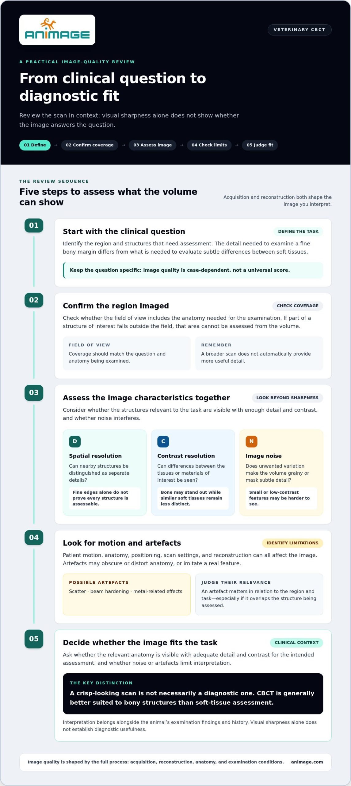
How to Assess Veterinary CBCT Image Quality in Practice
A repeatable review starts with the purpose of the scan, not a general impression of whether it looks clear. The sequence below is educational, not a clinical protocol. It helps organize observations, while the interpreting veterinary professional determines whether an examination is clinically adequate and how its findings should be understood.
A concise image-review sequence
- Confirm the question and region. Identify the clinical question, the anatomical region imaged and the specific structures that need assessment. Check that the relevant anatomy is included. A scan can only answer a question if the target is captured and displayed with sufficient detail for that task.
- Check coverage and presentation. Review whether the scan includes the region of interest and whether positioning gives a useful view of the anatomy. Look for signs of motion, uneven visibility, noise or artefacts. Note where they occur and whether they overlap structures central to the examination.
- Judge the limitation against the question. Ask whether the relevant anatomy can still be assessed despite any image limitation. An artefact elsewhere in the volume may have little effect on the task; one overlying the structure of interest may restrict interpretation.
This sequence separates observation from conclusion. First describe what the volume shows and where visibility is limited. Then consider whether that limitation affects the specific assessment. Avoid treating apparent sharpness, a single technical measure or the presence of an artefact as a complete verdict on image quality.
When image quality and modality choice intersect
Image quality and modality suitability are related, but they aren’t the same. A scan may be technically clear yet not provide the type of information needed for a particular question. CBCT is generally suited to bony structures, while its lower contrast resolution can limit soft-tissue assessment compared with conventional CT. The appropriate modality depends on the diagnostic task and clinical context, not a universal ranking.
For example, a question focused on fine bony detail calls for different image characteristics than one requiring tissue contrast. Review the structures that matter, the limitations that affect them and whether the chosen examination can address the question. For broader context, see veterinary CBCT uses, strengths, and limits.
Clinical adequacy isn’t established by appearance alone. The veterinary professional interpreting the study brings the examination findings, history and diagnostic purpose together with the image. This framework supports a more consistent discussion of veterinary CBCT image quality without replacing that judgment.
To explore a veterinary imaging platform that combines CBCT, digital radiography and fluoroscopy, learn about FIDEX+.
Where FIDEX+ Fits in a Veterinary CBCT Imaging Workflow
Image quality assessment helps clarify what a scan can show. The next consideration is how the imaging platform fits into the clinic’s workflow. FIDEX+ is Animage’s veterinary multi-modality imaging system, combining cone-beam CT (CBCT), digital radiography and fluoroscopy in one compact device. It brings these imaging methods together without requiring dedicated CT-room construction. This integration does not establish a particular image-quality outcome or determine which method is appropriate for an individual examination.
Each modality produces images in a different way and serves a different role. The choice still depends on the clinical question, the area being examined and the information the veterinary team needs. Platform integration can inform operational planning, but it doesn’t replace clinical selection of an imaging method or interpretation by a qualified professional.
Considering image quality within a multi-modality platform
Image quality remains specific to the modality and examination. Assess a CBCT scan in relation to the structures and question relevant to that scan. Digital radiography and fluoroscopy have their own imaging characteristics and uses. Having all three in one system doesn’t make their images interchangeable or make one modality the right choice for every task.
For workflow planning, a multi-modality system brings these imaging functions together in one equipment context. Consider how each method fits the clinic’s intended imaging workflow, while clinical judgment continues to guide method selection and assessment. The platform is a tool, not a substitute for that judgment.
Space and equipment planning context
FIDEX+ is a compact veterinary imaging system that doesn’t require dedicated CT-room construction. This can inform how a clinic considers equipment within its facilities and plans its use alongside other clinical operations. The system’s compact design doesn’t mean every existing space will suit every installation, so facility layout and intended workflow remain relevant planning factors.
Planning still involves considering the system in the context of the clinic’s facility and intended workflow. The educational framework in this article remains useful: define the examination question, assess the relevant images and their limitations, and leave clinical adequacy to the interpreting veterinary professional. Integration changes the equipment context, not the need to assess veterinary CBCT image quality according to the examination.
For clinics exploring an integrated veterinary imaging platform, FIDEX+ brings CBCT, digital radiography and fluoroscopy together in one compact system. Explore FIDEX+ to consider how its multi-modality approach could fit your imaging workflow.
Make Image Assessment Part of the Next Workflow Decision
Use the next imaging review as an opportunity to make the diagnostic question explicit before acquisition. A shared understanding of what needs to be assessed helps the veterinary team discuss image limitations in practical terms and keep technical appearance separate from clinical significance. The interpreting professional remains responsible for deciding whether a study is adequate for its purpose.
For clinics considering imaging equipment, review platform options alongside their intended use and facility needs. This keeps veterinary CBCT image quality in context: one part of a broader imaging decision, not a standalone promise of diagnostic success.
Explore the FIDEX+ veterinary imaging platform and consider how it could fit your clinic’s imaging plans. A clear question and a considered workflow are a practical place to start.
Frequently Asked Questions
What factors affect veterinary CBCT image quality?
Veterinary CBCT image quality reflects the full imaging process, including acquisition, patient positioning, exposure, reconstruction and the anatomy being examined. Noise or artefacts can also affect visibility. These factors interact: a technically well-sampled image may still be difficult to interpret if movement blurs a small structure. Review the scan in the context of its intended use rather than relying on one technical setting or a general impression of sharpness.
Are smaller voxels always better for veterinary CBCT?
No. A smaller voxel represents finer sampling, but it doesn’t guarantee that the scan will show more useful detail. The visible result also depends on factors such as noise, motion and the imaging system. The needed level of detail depends on the task: an examination focused on a small structure may have different requirements from one assessing a larger area. Voxel size is one part of image assessment, not a standalone quality rating.
Can motion make a veterinary CBCT scan difficult to interpret?
Yes. Movement during image acquisition can blur edges or make structures appear doubled or less distinct. This may matter most when the question concerns a small feature or a precise boundary. Review whether the affected anatomy remains assessable, rather than assuming every sign of motion invalidates the entire volume. Decisions about patient preparation and handling belong to the veterinary team and depend on the individual patient and examination.
What does field of view mean in a CBCT examination?
Field of view, or FOV, describes the portion of anatomy included in the scan. For example, an examination targeting a particular jaw region needs to include the structures relevant to that question. If an area of interest falls outside the captured volume, it can’t be evaluated from that scan. A wider field isn’t automatically preferable; the useful coverage depends on the purpose of the examination.
Does CBCT show soft tissue as clearly as conventional CT?
Generally, no. CBCT typically has lower contrast resolution than conventional CT, which can make differences between soft tissues harder to distinguish. This doesn’t mean CBCT is unsuitable for every examination. It means the veterinary team should match the modality to the diagnostic question and the tissues of interest. If the task depends on subtle soft-tissue contrast, that limitation should be considered when planning and interpreting imaging.
How can a veterinary team tell whether a CBCT image is diagnostically useful?
Start by stating the clinical question and identifying the structures needed to address it. Then check whether those structures are included and sufficiently visible, considering any motion, noise or artefact that overlaps them. A scan can have a limitation without that limitation preventing assessment. The interpreting veterinary professional determines clinical adequacy, using the images alongside the patient’s history and examination findings.
Can metal or other dense materials create artefacts on CBCT images?
Yes. Metal or other dense materials can contribute to beam-hardening or streak artefacts, which may distort the appearance of nearby anatomy or make it harder to assess. The effect varies with the material, its position and the structures around it. A streak doesn’t automatically indicate disease, nor does every artefact have the same cause. The key is to note whether it obscures anatomy relevant to the examination.