A dental radiograph can leave important anatomy hidden. In a veterinary CBCT system comparison, start with what CBCT does: cone-beam computed tomography rotates a cone-shaped X-ray beam around an animal and reconstructs images from multiple angles into a three-dimensional view. This can reveal details of teeth, roots, jawbones and surrounding structures that two-dimensional images may not show.
Image quality is only one consideration. Scan time, radiation exposure, clinical use and the practical value of the equipment all affect whether a system fits a veterinary setting. CBCT scans are typically completed within minutes, and veterinary CBCT typically requires a lower radiation dose than conventional CT.
This article explains how CBCT works and where its 3D imaging can support assessment of dental and maxillofacial conditions. It also compares system value through imaging capability, workflow, scan time and exposure, then considers how purchase costs depend on system configuration and intended use. The aim is a clear basis for judging which capabilities matter in a clinic, referral hospital or research setting.
Key Takeaways
- CBCT reconstructs a rotating cone-beam X-ray scan into a 3D view, helping reveal dental and maxillofacial detail that can be obscured on 2D radiographs.
- Assess cost-effectiveness against the cases a system can support, its imaging capabilities and how it fits into clinical workflow, not purchase cost alone.
- Veterinary CBCT scans are typically completed within minutes; scan duration is only one part of the practical workflow.
- Veterinary CBCT typically requires a lower radiation dose than conventional CT, while patient anatomy and the region being imaged shape the examination.
- FIDEX+ combines veterinary CBCT, digital radiography and fluoroscopy in one compact imaging platform.
3D imaging of the oral and maxillofacial region
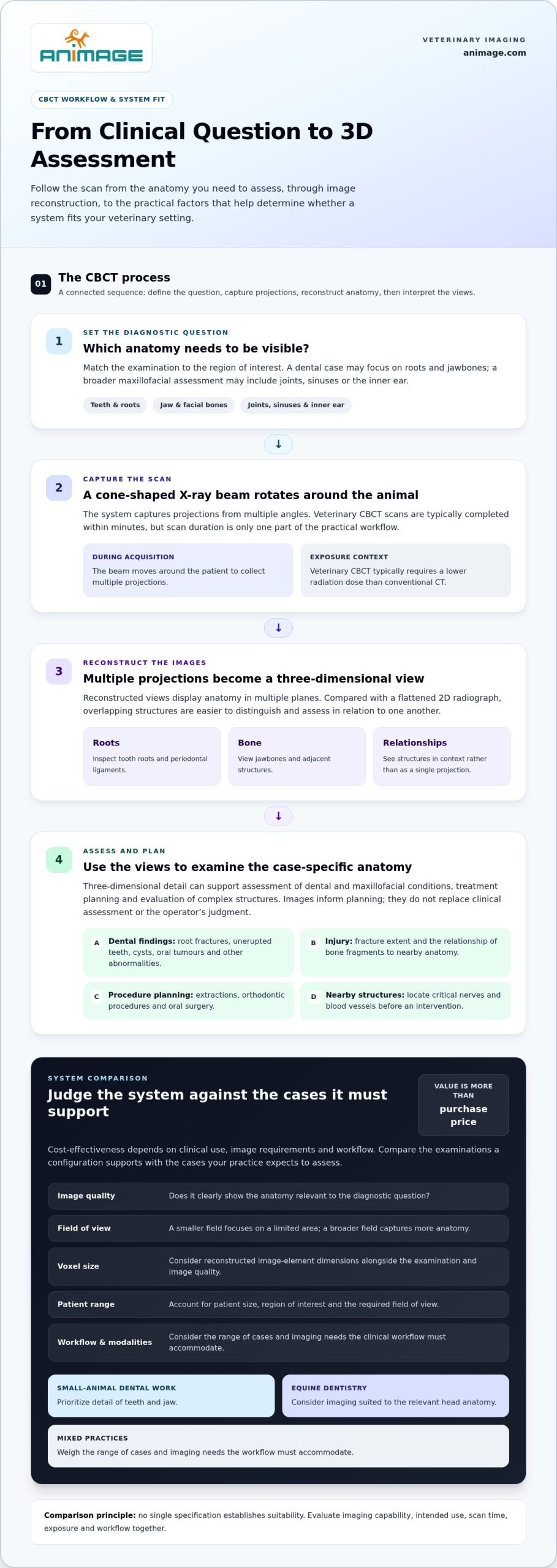
Veterinary cone-beam computed tomography uses a cone-shaped X-ray beam that rotates around the animal. The system captures projections from multiple angles and reconstructs them into a three-dimensional representation. Unlike a two-dimensional dental radiograph, this view displays anatomy in multiple planes, making overlapping structures easier to distinguish. Teeth, roots, jawbones and nearby tissues can be assessed in relation to one another, rather than as a flattened projection.
That difference matters when a dental problem lies within or behind surrounding bone. CBCT can show tooth roots, periodontal ligaments and adjacent bone in detail. For example, a suspected root fracture can be examined across reconstructed views, rather than from a single radiographic direction. Images can also help locate unerupted teeth and characterize cysts, oral tumours or other abnormalities. A veterinary CBCT system comparison should consider whether each system clearly images the anatomy relevant to the diagnostic question.
The oral and maxillofacial region extends beyond the teeth. CBCT can depict the temporomandibular joints, sinuses and inner ear, helping assess abnormalities in these structures. In trauma cases, three-dimensional views can show the extent of a fracture and how bone fragments relate to nearby anatomy. This information can support treatment planning and help clinicians evaluate complex structures that may be difficult to interpret on conventional dental radiographs alone.
- Tooth roots and surrounding bone: assess root structure, periodontal ligaments and adjacent bone for dental disease or injury.
- Jaw and facial bones: map fractures, trauma and the extent of bone involvement.
- Abnormal growths: examine oral tumours, cysts and other pathologies in relation to nearby anatomy.
- Joint and sinus regions: investigate the temporomandibular joints, sinuses and inner ear.
Three-dimensional anatomy can also support planning for extractions, orthodontic procedures and oral surgery. A reconstructed view shows where a tooth root lies relative to surrounding bone and can help locate critical nerves and blood vessels before an intervention. This spatial information may guide a more precise approach and help clinicians avoid important structures. Images inform planning, but do not replace clinical assessment or the operator’s judgment.
When comparing systems, focus on the intended region of interest, reconstructed detail and clinical workflow. A dental examination may prioritize fine structures around roots and jawbones. A broader maxillofacial assessment may require views of joints, sinuses or the inner ear. The useful question is not simply whether a scanner produces 3D images. It is whether those images show the anatomy needed to answer the case-specific diagnostic question.
For dental assessment, veterinary cone-beam CT provides high-resolution three-dimensional images of the oral and maxillofacial region in animals. Detailed views of teeth, jawbones and surrounding structures can help diagnose dental conditions. CBCT is the gold standard for dental imaging in animals, particularly in complex cases involving root fractures, unerupted teeth and jaw tumours. It can also help detect and characterize oral tumours, cysts and other pathologies, showing their relationship to nearby anatomy.
In cases of oral trauma or fractures, CBCT can show the extent of the injury and help guide treatment. Its detailed three-dimensional representation can also help veterinarians plan surgical procedures with greater precision. These capabilities are useful when the clinical question requires a clear view of both the affected structure and surrounding anatomy.
CBCT cost-effectiveness
Cost-effectiveness depends on clinical use, image requirements and workflow, not purchase price alone. A system used for dental cases must show the relevant anatomy clearly; a broader service may benefit from additional imaging functions. Compare the examinations each configuration supports with the cases your practice expects to assess. This grounds the investment in clinical needs rather than a single specification.
When assessing image quality, field of view and voxel size, compare the region captured with the detail the diagnostic task requires. A smaller field of view focuses imaging on a limited area, while a broader one captures more anatomy. Voxel size describes the dimensions of reconstructed image elements and should be considered alongside the intended examination and image quality. Together, these criteria help distinguish systems without treating one specification as a complete measure of performance.
Which systems suit small animals, equine dentistry and mixed practices? The fit depends on patient size, the region of interest and the required field of view. Small-animal dental work prioritizes tooth and jaw detail; equine dentistry requires imaging suited to the relevant head anatomy. Mixed practices should weigh the range of cases and imaging needs their workflow must accommodate.
A specification list is useful only in context. Compare veterinary CBCT systems by image quality, field of view, voxel size, intended patient range and supported modalities. For a named model, use its technical documentation to assess those details. In Poland, evaluate systems designed for veterinary imaging against the same criteria, rather than assuming that a brand name alone establishes suitability.
Total ownership cost includes more than equipment acquisition. Budget planning should account for installation, staff training, software, routine maintenance and service arrangements. Warranty terms and service options differ by supplier and agreement, so include them in the whole-life comparison. Price depends on the system configuration and intended application, and is provided after a quote request; imaging capabilities, included modalities and installation requirements shape the cost.
Room, power and shielding requirements depend on the selected system and installation site. They affect preparation and operating costs, so include them in early planning. Training and image interpretation support also influence practical value: software must fit the clinic’s workflow, and staff need to use and interpret reconstructed images competently. Compare what each system includes, then weigh those elements against expected use. A system with broader capabilities may serve more clinical tasks, but value comes from capabilities the practice can use, not specifications it does not need.
CBCT scan times
Veterinary CBCT scans are typically completed within minutes. That describes image acquisition, not necessarily the full appointment. Positioning the patient, selecting the region of interest, setting up the examination and reviewing reconstructed images all contribute to the workflow. A focused dental scan and a broader skull examination may require different preparation. Compare systems by the complete clinical process, not acquisition time alone.
The scan begins with positioning. The team aligns the patient with the field of view, the area the scan will capture. Accurate positioning matters: a dental examination focused on one jaw region differs from imaging the wider maxillofacial area. The operator then selects a scan protocol suited to the clinical question. The cone-shaped X-ray beam rotates around the patient, capturing projections that software reconstructs into a three-dimensional image.
Acquisition time can depend on the selected field of view and imaging protocol. A limited region may need a different scan setup from a broader anatomical assessment. Patient movement during acquisition can also affect image quality, making preparation and positioning practical factors in a time comparison. A short scan is useful only if it captures the anatomy needed for interpretation. Speed alone does not establish diagnostic value.
- Preparation: position the patient and align the intended anatomy within the field of view.
- Acquisition: run the selected CBCT protocol while the system captures projections.
- Reconstruction: generate the three-dimensional dataset for review.
- Interpretation: assess whether the images answer the clinical question, such as locating a tooth root or evaluating jaw anatomy.
These stages explain why two systems with similar acquisition times may produce different appointment workflows. Interface design, protocol selection and image reconstruction affect how efficiently staff move from setup to review. For a referral hospital, predictable transitions between examinations may matter more than a brief scan in isolation. In a practice with occasional CBCT cases, ease of selecting the correct region may carry greater weight.
When comparing veterinary CBCT systems, ask what the stated scan time covers: acquisition only or another defined part of the workflow. Then consider the examination type, positioning demands and image review process together. A system used for dental cases should support a clear workflow from patient alignment to reconstructed images. The relevant measure is not simply how quickly the scanner rotates, but how efficiently the team obtains usable images for the intended assessment.
Radiation dose and patient anatomy
Assess radiation exposure alongside the anatomy a scan needs to show. Veterinary CBCT typically requires a lower radiation dose than conventional CT, but not every CBCT examination is identical. The selected protocol, field of view and patient positioning affect how the system captures the target region. A focused tooth scan and a broader skull examination answer different clinical questions.
Patient anatomy guides the scan area. A small animal’s dental examination may focus on teeth, roots and adjacent jawbone. A larger anatomical region may be needed to assess facial trauma or sinus abnormalities. Selecting a field of view that matches the clinical question keeps the examination focused on relevant anatomy. A field that is too narrow may omit structures needed for assessment; an unnecessarily broad one captures more anatomy than the question requires.
Positioning matters too. The patient must be aligned so the intended region falls within the scan area. An examination focused on a jaw, for example, needs different alignment from one assessing the wider maxillofacial region. Movement during image acquisition can affect the resulting dataset. Preparation and positioning therefore contribute to image quality, which matters when weighing the benefit of an exposure.
- Clinical question: identify whether the examination targets a tooth, jawbone or broader skull region.
- Field of view: match the captured area to the anatomy required for assessment.
- Patient alignment: position the target anatomy within the selected scan area.
- Image review: assess whether reconstructed views show the structures relevant to the case.
These criteria help compare systems without relying on a dose figure in isolation. The same scanner may use different protocols for different anatomical targets. A meaningful comparison considers the examination type and region together. A dental protocol and a broader skull protocol, for example, should not be treated as equivalent simply because both use CBCT.
In practice, assess dose information for the intended examination, not an unrelated example. Consider whether the system can capture the region of interest with appropriate image detail and whether its workflow supports accurate positioning. Veterinary CBCT’s typically lower dose than conventional CT is relevant, but does not replace protocol selection based on patient anatomy. A useful comparison connects exposure, scan area and the diagnostic question.
Resolution and precision also matter when assessing the anatomy a scan needs to show. Veterinary CBCT offers higher resolution and precision than traditional dental radiographs for examining dental and anatomical structures. This matters when the clinical question depends on identifying details within the selected region, rather than simply capturing a broad view. Consider the value of that detail alongside the examination target and patient positioning.
Veterinary cone-beam CT can help veterinarians locate anatomical structures and abnormalities when planning minimally invasive procedures. CBCT scans are typically completed within minutes, helping minimise patient discomfort and stress. Together, image detail and scan duration relate to the practical demands of assessing patient anatomy: the examination should provide information relevant to the procedure while keeping the patient’s experience in view.
Radiation exposure compared with conventional CT
Veterinary CBCT typically requires a lower radiation dose than conventional CT, but the modalities differ in image formation and clinical use. CBCT rotates a cone-shaped X-ray beam around the patient and reconstructs projections into a three-dimensional dataset. Conventional CT uses a different acquisition approach. Dose comparisons matter, but do not establish which examination best answers a particular question.
What are CBCT’s advantages and limitations compared with veterinary CT and dental radiography? CBCT provides high-resolution three-dimensional views of teeth, roots and jawbones, while dental radiographs show anatomy in two dimensions. Compared with conventional CT, CBCT typically requires a lower radiation dose. Each modality should be matched to the anatomy and diagnostic task.
For a suspected tooth-root problem, a dental radiograph may provide a useful projection, but overlapping structures can obscure detail. CBCT adds reconstructed views across multiple planes, helping assess the root and surrounding bone spatially. That advantage depends on the question being examined. A three-dimensional dataset is not automatically more useful if the clinical question concerns anatomy outside the captured region.
Conventional CT remains a distinct option for veterinary imaging. The comparison is not simply dose versus dose: it also includes the anatomy to assess, the required field of view and the information each modality can provide. A focused dental assessment and a broader skull examination, for example, may require different imaging considerations. The selected modality should fit the region of interest.
- Dental radiography: provides two-dimensional projections for assessing dental structures.
- Veterinary CBCT: reconstructs high-resolution three-dimensional images, particularly useful for oral and maxillofacial anatomy.
- Conventional CT: offers a separate acquisition approach for cases requiring a different view or examination scope.
CBCT’s lower typical dose than conventional CT is one factor in system evaluation, not a standalone measure of suitability. Captured anatomy, image detail and scan protocol matter too. For a jaw fracture, the clinician needs views that show its extent and relationship to adjacent structures. For a dental concern, the focus may instead be roots and surrounding bone.
Compare modalities using the same clinical question and anatomical target. Consider whether the examination needs a two-dimensional projection or reconstructed 3D views, then assess the relevant field of view and image detail. This keeps the comparison grounded in clinical value rather than a single dose claim. CBCT can add useful anatomical information, but it is not a universal replacement for dental radiography or conventional CT.
Animage: FIDEX+
Animage’s FIDEX+ brings veterinary cone-beam CT, digital radiography and fluoroscopy together in one compact imaging platform. Designed specifically for animals, it offers clinics, referral hospitals and research institutions a multi-modality option when evaluating imaging equipment for their workflow.
The combination of modalities is relevant for practices comparing a dedicated CBCT system with a platform that also includes radiography and fluoroscopy. Its practical value depends on the examinations your team performs and how those imaging methods fit into daily use. Evaluate FIDEX+ against your clinical setting, imaging priorities and intended cases.
Contact Animage to discuss how FIDEX+ fits your veterinary imaging requirements.
Turn the comparison into a practical decision
Move from specifications to a clear picture of how imaging should function in your practice. Identify the cases the equipment needs to support, who will use it and how examinations should fit into the existing workflow. This gives your team a shared basis for evaluating capabilities, implementation priorities and long-term clinical value.
A veterinary CBCT system comparison is most useful when it connects each technical requirement to an operational need. Separate essential functions from capabilities that would be used only occasionally. Then consider whether a multi-modality platform belongs in the evaluation. FIDEX+ is Animage’s veterinary imaging platform for teams considering that approach.
Use those priorities to frame the next discussion and clarify what matters for your setting. Contact Animage to discuss CBCT and explore whether FIDEX+ aligns with your imaging requirements. A focused decision starts with the cases your practice needs to serve.
Frequently Asked Questions
What is CBCT?
CBCT, or cone-beam computed tomography, is an X-ray imaging method that produces a three-dimensional dataset for veterinary assessment. It can help show how structures relate within a region, rather than relying only on a flat projection. For example, images of the eye socket may help assess a suspected foreign object or fracture and inform planning for an ophthalmic procedure. The clinical question determines which anatomy needs evaluation.
How do veterinary CBCT systems compare in image quality, field of view, and voxel size?
Compare image quality and voxel size with the detail needed for the intended examination. A smaller field of view focuses on a limited area, while a broader one captures more anatomy. No single specification determines performance on its own.
What are the main veterinary CBCT system brands and models, and their specifications?
FIDEX+ combines veterinary CBCT, digital radiography and fluoroscopy in one compact platform. The article does not provide comparative specifications for other models.