Veterinary CBCT can suit dental, maxillofacial, orthopedic and other detailed bone imaging, while conventional CT offers broader flexibility for soft-tissue and body-cavity studies. If you’re deciding how to choose veterinary CBCT, start with the cases you need to image, not a scanner’s headline features. The practical difference is that each system supports different diagnostic priorities.
That’s why a compact footprint or detailed 3D images shouldn’t decide the purchase alone. Patient size, positioning, radiation safety, room requirements and the full cost of ownership also shape whether a system fits your clinic’s workflow. A mismatch can limit the studies you can perform or create avoidable operational demands.
This guide compares CBCT with conventional CT for clinical use, image requirements, mobility, facility planning and costs in Poland. You’ll learn which factors matter for your caseload, where each system has limitations, and how to weigh current needs against future flexibility.
Key Takeaways
- Match the scanner to your expected case mix: CBCT suits detailed bone-focused imaging, while conventional CT supports broader soft-tissue and body-cavity studies.
- Plan radiation protection and room requirements around the system and applicable Polish requirements before committing to an installation.
- Assess patient size, positioning and scanner geometry together; a compact footprint alone doesn’t establish whether a system fits your caseload.
- Compare fixed and mobile configurations by considering where patients are scanned and how the equipment will fit into daily workflows.
- Build a whole-ownership view of costs, including installation, maintenance and software, then assess whether FIDEX+’s combined imaging modalities align with your clinic’s needs.
Radiation safety and facility requirements
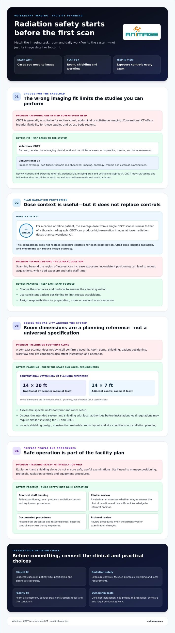
Radiation planning starts with the room, workflow and species being scanned. Veterinary cone-beam computed tomography (CBCT) can produce high-resolution images at lower radiation doses than conventional CT. For a canine or feline patient, the average dose from a single CBCT scan is similar to that of a thoracic radiograph. This comparison helps put exposure in context, but does not replace exposure controls for each examination.
What training and staffing are needed to operate veterinary CBCT safely? Staff need practical training in patient positioning, scan protocols, radiation controls and equipment procedures. A veterinarian must assess whether the images answer the clinical question and have sufficient knowledge to interpret the findings.
What are the limitations, risks, and contraindications of veterinary CBCT in animals? CBCT uses ionising radiation, and movement can reduce image accuracy. It is generally unsuitable for routine thoracic, abdominal or soft-tissue imaging, where conventional CT can provide better tissue contrast and greater flexibility.
Use a pre-installation review to turn radiation-safety requirements into facility decisions. The International Atomic Energy Agency’s veterinary radiation-safety guidance covers facility design and protection for workers and the public. The American College of Veterinary Radiology explains the CT process and the need for conscientious radiation-safety practices in veterinary CT. Before installation, discuss the intended system and shielding with local authorities. Local regulations may require similar shielding for CT and CBCT.
Room planning also affects installation costs. A traditional veterinary CT scanner room should be at least 14x20 feet, with an adjacent control room of at least 14x7 feet. These dimensions are planning references for conventional CT, not universal CBCT specifications. Shielding design, construction materials, room layout and site conditions shape the final project cost. Include these items in the budget alongside installation, equipment and any required building work.
| System option | Typical imaging focus | Facility planning | Key safety consideration |
|---|---|---|---|
| Veterinary CBCT | High-detail bone imaging | Assess the specific unit’s footprint and room setup | Set exposure controls and positioning procedures |
| Conventional CT | Broader tissue and body-region imaging | Plan scanner room and adjacent control room | Apply radiation protection to the full workflow |
Reduce radiation dose by choosing a scan area and protocol suited to the clinical question. Avoid imaging beyond the region of interest, and use consistent positioning to limit repeat acquisitions. A focused dental examination, for example, needs different coverage from a study of a larger anatomical region. Repeat scans increase exposure and take staff time.
Build safety into daily operation, not just facility design. Assign staff responsibilities for patient preparation, room access and scan execution. Document local procedures, keep the control area clear during exposures, and review protocols when the patient type or examination changes. When deciding how to choose veterinary CBCT, weigh the imaging tasks against room design, shielding requirements, dose management and operating costs.
Why choose Cone Beam Computed Tomography for veterinary radiography?
Choose based on clinical workload, not image detail alone. Cone-beam CT suits focused imaging needs; conventional CT supports broader diagnostic coverage.
Clinical Caseload and Applications
Veterinary CBCT is suited to dental, oral and maxillofacial imaging, orthopaedics, trauma, and detailed bone assessment. Its 3D images can help identify dental anomalies, fractures and joint disorders, supporting diagnosis and treatment planning. This makes it relevant to clinics whose routine referrals concentrate on those cases, rather than a broad mix of body systems.
Conventional multislice CT is the more flexible option for a broad caseload. It supports soft-tissue studies, thoracic and abdominal imaging, oncology, trauma and contrast examinations. CBCT is generally unsuitable for routine imaging of the chest, abdomen or soft tissue. Review the cases your clinic performs and expects to add, then compare them with each system’s diagnostic strengths.
Workload matters as much as case type. A clinic handling frequent dental referrals may benefit from a system that supports detailed oral imaging within its existing workflow. A practice expecting varied studies across body regions may need conventional CT’s broader diagnostic coverage. Consider how referrals, image interpretation and follow-up planning fit into daily operations.
Patient mix also guides the decision. CBCT can be useful for small mammals and exotic animals, as well as canine and feline dental or maxillofacial cases. Consider whether the system’s imaging area and positioning approach suit the examinations your practice typically performs. For patients needing broad soft-tissue assessment, conventional CT offers greater flexibility.
Clinical and Business Benefits
CBCT’s main clinical advantage is detailed visualization of complex bone structures. A cone-shaped X-ray beam and two-dimensional detector generate 3D images, providing high spatial resolution for focused examinations. Faster scan times can reduce the need for prolonged animal sedation. These features support targeted diagnostic work, but they don’t make CBCT a substitute for every CT examination.
Some CBCT systems have a compact footprint, which can make them easier to integrate into clinics with limited space. Their value depends on whether they suit the caseload and support useful clinical services without displacing essential room functions. Estimate staff workload, patient preparation, image review and referral demand alongside scanner fit. A compact unit helps only when the entire workflow works.
A practical approach to how to choose veterinary CBCT is to map common examinations to imaging needs, then assess space and workflow. Explore integrated veterinary imaging workflows as one way to consider how multiple imaging modalities may fit within a clinic.
Patient size, scanner geometry, and positioning
Patient dimensions and the target anatomy determine whether a scanner can capture the required diagnostic field. Compare the largest animals you routinely image with the smallest patients needing precise positioning. A standard veterinary CT scanner bore may have a 70 cm diameter for larger animals, such as farm animals. That figure illustrates conventional CT geometry, not a universal CBCT dimension.
For CBCT, compare the field of view with the anatomy you need to image. A dental examination may require a focused view of the jaw, while a larger orthopedic study needs coverage of the relevant joint and surrounding structures. Field of view, voxel size and image resolution describe related but distinct capabilities.
How should you compare veterinary CBCT field of view, voxel size, and image resolution? Match the field of view to the anatomy, then compare voxel size and resolution with the detail required for that study. A smaller voxel value alone does not establish better diagnostic performance for every patient or examination.
Positioning determines whether the planned region fits within the capture area. Before selecting a scanner, map the patient’s posture and target anatomy for common studies, such as standing limb imaging or dental positioning. Consider access around the patient, restraint workflow and how staff can maintain consistent positioning. These requirements vary by patient size and examination type.
Scanner geometry also affects the range of patients and studies a clinic can accommodate. A compact system may suit focused imaging in a smaller clinical space, while larger patients or broader examinations may need a different configuration. Compare the usable imaging area and patient access with the animals in your caseload, not just the scanner’s external footprint.
Mobile CT is another option for practices serving patients at farms. Mobile CT scanners are installed in trailers, enabling scanning at locations such as farms. A fixed scanner brings imaging into the clinic workflow. Consider where patients are located, how they reach the scanner and whether moving the equipment or the patient best suits your service model.
Compare veterinary CBCT scanner models by clinical coverage, geometry and positioning workflow, not just by model name. Some suit dental and maxillofacial work; others may support additional bone-focused examinations. Conventional CT offers broader coverage for soft tissue and body regions. Match the modality to the studies your practice intends to perform.
Compare systems against a list of expected patient sizes, examination types, positioning needs and image-sharing steps. Then assess each scanner configuration against that list. This ties technical specifications to real cases and helps reveal where a system’s geometry or software workflow could limit routine use.
Scanner mobility and compact design
Mobility affects where imaging can take place; compact design affects how a scanner fits onsite. Veterinary CBCT systems can have a compact footprint, which may simplify integration into smaller practices. Conventional CT can support broader diagnostic coverage, but its room and workflow requirements differ. Compare the equipment’s footprint with patient access, staff movement and the examinations your clinic performs.
How does veterinary CBCT compare with CT, dental radiography, and MRI? CBCT produces three-dimensional images suited to detailed bone-focused studies, while conventional CT supports broader imaging, including soft tissue and body regions. Dental radiography provides targeted views, and MRI is a separate imaging modality, not a type of computed tomography.
A compact scanner is useful only if the animal can be positioned for the required study. For example, a clinic focused on dental imaging needs practical access for oral positioning and patient preparation. A system intended for additional orthopedic or trauma imaging must also accommodate those workflows. Assess the full setup, including positioning space, staff access and movement around the patient.
Fixed and mobile systems address different location needs. Mobile CT scanners are installed in trailers to scan patients at locations such as farms. This can bring imaging closer to animals that are difficult to transport. A fixed clinic scanner places examinations within the hospital workflow, where staff can prepare patients and coordinate imaging with other clinical activities.
Compare mobility by mapping where scans will take place and who needs access to the equipment. A farm-based service may benefit from trailer installation and travel logistics. A referral hospital may prioritize access within the facility and integration with its diagnostic workflow. Patient transport, staff time and examination location all influence whether mobile or fixed imaging is the practical configuration.
The imaging modality matters more than a compact footprint alone. CBCT can deliver high spatial resolution for complex bone structures, including dental and maxillofacial anatomy. Conventional CT is generally more suitable for broad caseloads, thoracic and abdominal studies, and soft-tissue assessment. In larger dogs and body regions such as the thorax, conventional CT usually provides better soft-tissue contrast and is less susceptible to motion artefacts.
Before choosing, compare sites and studies. List where imaging must take place, which patients need access, and the anatomical regions commonly examined. Then assess fixed versus trailer-based CT and compact CBCT against those needs. Choose the configuration that supports the required studies and fits the clinic’s actual workflow, not just its available floor space.
Veterinary CBCT system costs and return on investment
Compare purchase, installation and operating costs over a five-year ownership period. A lower acquisition cost matters only if the scanner suits the clinic’s imaging workload.
Purchase and Installation Costs
Purchase and installation make up the initial investment, but costs vary by system and site. A veterinary CT scanner’s price depends on its type and whether it is new or refurbished. Installation planning should include room preparation, shielding, electrical requirements and workflow integration. For Polish clinics, assess these items in zł and include applicable site work in the project budget.
CBCT systems may cost less to purchase than conventional CT, but compare full project estimates. The price depends on the case and is provided after a quote request. Cost factors include scanner configuration, installation work, room modifications and technical integration. Keep equipment costs separate from unrelated clinical fees when modelling the investment.
Maintenance and Replacement Costs
Operating expenses distinguish a scanner’s purchase price from its longer-term financial load. CBCT scanners can have up to 70% lower ongoing maintenance and replacement costs than traditional fan-beam CT scanners. Compare service requirements, software costs and replacement parts over the same ownership period. Use annual estimates in zł, and separate recurring expenses from occasional component replacement.
Pay particular attention to the X-ray tube. Replacing a conventional CT tube can be a major ownership expense, while CBCT tubes typically cost less to replace. Ask about the expected replacement interval and include the tube in lifecycle planning rather than treating it as an unexpected repair. Also account for scanner downtime, which can affect scheduled imaging and referrals.
Return on Investment
Return on investment depends on the clinical cases a system can support and its total cost of ownership. CBCT focused on detailed bone imaging may suit a clinic handling dental studies, fractures and other complex structures. Conventional CT can serve a broader caseload, including soft-tissue examinations. Estimate utilization from your actual case mix, not assumed demand.
Build a five-year comparison that includes acquisition, installation, maintenance, software and replacement components. Then assess how the scanner could affect referral handling, imaging capacity and the range of studies performed onsite. This gives a practical view of ownership, not a promise of payback. A system that cannot support the clinic’s required examinations may offer less value even if its running costs are lower.
Use your projected case mix and ownership assumptions to compare imaging configurations. Request a project quote to discuss system costs and installation factors for your clinic.
Animage: FIDEX+
FIDEX+ combines veterinary cone-beam CT, digital radiography and fluoroscopy in one compact imaging platform. For clinics assessing an integrated imaging setup, it brings three modalities together in a single device. It may suit practices whose caseload includes detailed bone imaging alongside radiographic and fluoroscopic examinations.
Use your case mix and facility plan to assess whether this approach fits. Consider the examinations you expect to perform, patient positioning, available room space and how staff will move between imaging tasks. FIDEX+ is designed specifically for animals and is sold to veterinary clinics, referral hospitals and research institutions. It is an option to consider when an integrated veterinary imaging platform matches your clinical and operational priorities.
Contact Animage to discuss how FIDEX+ could fit your imaging requirements.
Turn Your Imaging Priorities Into a Clear Decision
Start by defining what a successful imaging setup needs to support in your clinic. List the examinations that matter most, the patients and staff who will use the system, and the operational constraints that shape daily work. This gives your team a practical basis for comparing options and weighing trade-offs before committing to an equipment plan.
The decision should reflect the clinic’s intended role, not just a scanner’s technical appeal. If your priorities point toward an integrated veterinary imaging platform, consider FIDEX+ from Animage. Use your requirements to guide a focused discussion about how its approach aligns with your practice’s needs.
Contact Animage about FIDEX+ and take the next step toward an imaging plan built around your clinical priorities.
Frequently Asked Questions
What veterinary CBCT specifications matter for small animal clinics?
Prioritize specifications that match the clinic’s most common examinations, such as dental, maxillofacial or orthopedic studies. Compare field of view and image detail using representative cases, including the smallest patient and the most complex target anatomy. Then assess how image review fits your reporting workflow. A specification matters clinically only if it supports the regions you need to examine and the decisions your team must make.
What room size, shielding, and electrical requirements does veterinary CBCT installation need?
Room dimensions and electrical requirements depend on the installed system and its configuration. Plan space for patient transfer, positioning, staff access and equipment operation, not just the scanner footprint. Local regulations may require similar radiation shielding for CT and CBCT systems. In Poland, include radiation-protection planning and site electrical capacity in pre-installation work, with requirements tailored to the equipment and premises.
What maintenance, warranty, and service contract should a veterinary CBCT buyer in Poland expect?
Compare written warranty and service terms rather than assuming every system has the same coverage. Market information indicates that most CT scanners include a one-year warranty, while CBCT systems often include several years of technical support with the initial purchase. Review what the agreement covers, including parts, labour, software support, response process and exclusions. Record renewal terms and responsibilities in the purchase documentation to support clear ownership planning.
How to estimate patient volume, operating costs, and return on investment for veterinary CBCT?
Start with a five-year ownership estimate, then count the clinic’s annual cases suited to CBCT, not all imaging appointments. Separate existing cases from new referrals, and estimate revenue using the clinic’s own pricing and expected workload. Subtract installation, maintenance, software and replacement costs to compare scenarios. Model a conservative case mix alongside the expected one, so the decision is based on realistic utilization rather than projected demand alone.