A radiograph isn’t finished when the exposure ends. Veterinary digital radiography is a complete image workflow: a detector captures the X-ray exposure, software processes and displays the image, and the clinical team reviews and stores it. The detector matters, but so do image handling and the steps around each examination.
Film, computed radiography (CR), and direct digital radiography (DR) all produce radiographic images, but they differ in how images are captured and made available for review. Digital workflows can reduce the wait associated with film processing, but image processing doesn’t replace sound positioning, exposure technique, or clinical interpretation. A displayed image still needs to be assessed in context.
This article explains how image acquisition and review work, compares film, CR, and DR, and outlines practical workflow benefits and limitations. It also covers what to evaluate when selecting a system, including how it fits into the clinic’s wider imaging process. FIDEX+ combines digital radiography with cone-beam CT and fluoroscopy in one veterinary imaging platform. Each examination should use the modality suited to its purpose.
Key Takeaways
- Trace how veterinary digital radiography turns an X-ray exposure into a displayed image, and learn what pixels and grayscale represent.
- Compare film, computed radiography, and digital radiography by image availability, processing steps, storage, sharing, and workflow.
- Use patient mix, examination types, room layout, and staff routines to assess how a system could fit your clinic.
- Include image review, storage, transfer, and access needs when planning an imaging workflow.
- Understand the distinct role of 2D radiography alongside cone-beam CT and fluoroscopy in an integrated imaging platform.
What Is Veterinary Digital Radiography, and What Happens During an X-Ray?
Veterinary digital radiography captures X-ray images with an electronic detector instead of film, then displays them on a computer for review. Image acquisition creates a representation of the patient’s anatomy, not a clinical diagnosis. Interpretation depends on image quality, the clinical question, and the veterinary professional’s assessment.
The workflow starts before exposure. The patient is positioned for the required view, and the X-ray beam is directed through the area of interest toward the detector. After exposure, the system converts the detector’s response into image data. Software processes and displays the data for review. Positioning, exposure, capture, processing, and interpretation are connected steps, but they are not interchangeable.
- Position the patient. Align the anatomy and detector for the intended view. Movement or poor positioning can limit what the image shows.
- Make the exposure. X-rays pass through the patient, and different tissues reduce the beam by different amounts.
- Capture the signal. The detector records the X-rays that reach it and converts their pattern into digital data.
- Process and display the image. Software maps the data to shades of grey and presents the image on a monitor.
- Review and interpret. A veterinary professional assesses the image in context, including whether the view is suitable for the clinical question.
How Does a Digital Veterinary X-Ray Become an Image?
Bone, soft tissue, air, and other structures attenuate X-rays to different degrees. The detector therefore receives a varied signal across its surface. The system translates these differences into image contrast, with areas appearing lighter or darker according to the displayed signal. Pixels are the small picture elements that make up the digital image, and each is assigned a grey value.
Capture and display are separate. Once the detector has recorded the exposure, software can adjust how the image is presented, such as its brightness or contrast, to support review. These controls change the displayed rendering, not the positioning or exposure. They can’t recover detail that wasn’t captured or make an unsuitable view diagnostic. The overview of Digital radiography also explains the main digital approaches, including computed and direct capture.
What Does Digital Change Compared With Film?
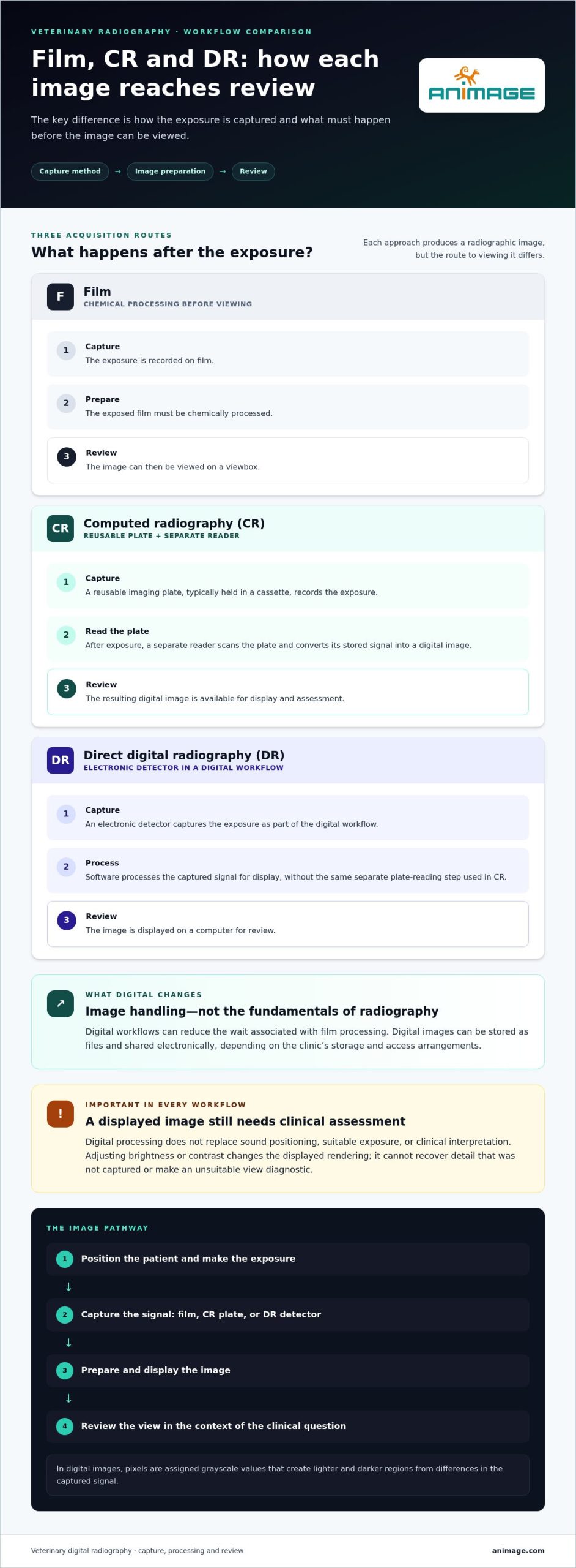
With film, the exposed cassette must be chemically processed before the image can be viewed on a viewbox. Digital radiography replaces that chemical step with computer-based image display. The image can then be stored as a digital file and shared electronically, depending on the clinic’s storage and access arrangements.
This changes how images move through a practice, not the fundamentals of radiographic technique. Digital capture doesn’t guarantee correct positioning, suitable exposure, or a diagnostic image. The team still needs to assess whether the view answers the clinical question and whether additional imaging is appropriate.
How Veterinary Digital Radiography Captures, Processes, and Displays Images
In veterinary digital radiography, an image passes through distinct stages. Separating them helps teams identify whether a limitation began during capture, arose during display, or relates to interpretation. In brief, a detector converts the pattern of X-rays it receives into pixel data, which software processes and displays as a digital radiograph.
- Exposure: The X-ray beam passes through the positioned patient. Tissues attenuate the beam by different amounts, so the detector receives a varied pattern.
- Detector response: The detector converts the received X-ray pattern into an electronic signal. How that signal is captured depends partly on whether the system uses computed radiography (CR) or direct digital radiography (DR).
- Image processing: Acquisition software translates the signal into image data and applies the system’s processing to prepare it for display. Processing follows capture; it does not replace it.
- Display: The software renders the data on a monitor as pixels, the small elements that make up the image. Each pixel is assigned a grayscale value, producing lighter and darker regions that represent differences in the captured signal.
- Storage and interpretation: The image may be saved and made available through the clinic’s digital workflow. A veterinary professional assesses what it shows and whether it is suitable. The software display itself isn’t a diagnosis.
Acquisition, processing, display, storage, and interpretation each serve a different purpose. For example, zooming in changes the view on screen, not the original patient positioning. The American College of Veterinary Radiology provides professional context for veterinary radiology and specialist interpretation.
What Is the Difference Between CR and DR?
CR uses a reusable imaging plate, typically held in a cassette. After exposure, a separate reader scans the plate and converts its stored signal into a digital image. DR captures the exposure with an electronic detector as part of a digital workflow, without the same plate-reading step. These are broad acquisition approaches, not identical equipment configurations. Hardware, handling, and workflow details vary between systems, so the labels alone don’t describe every operational difference.
What Can Image Processing Change?
Depending on the system, the reviewer may adjust brightness and contrast, zoom into a region, or use measurement tools. These functions can help make captured anatomy easier to examine. They cannot restore anatomical information that wasn’t recorded or correct motion and positioning problems after exposure. Image quality still depends on appropriate positioning and exposure technique, as well as the detector and acquisition workflow.
Plan for both image acquisition and the way staff review and access images. FIDEX+ combines digital radiography with cone-beam CT and fluoroscopy in one veterinary imaging platform. When assessing an integrated veterinary imaging platform, consider how its modalities fit examination pathways. Not every case needs more than one modality.
Digital Radiography Versus Film and CR: What Changes in Practice?
The practical difference between film, computed radiography (CR), and direct digital radiography (DR) is how an image moves from exposure to review. Film requires chemical processing. CR adds a separate plate-reading step. DR captures the exposure through an electronic detector in a digital workflow. Exact handling and system capabilities vary, so these categories describe broad approaches rather than identical setups.
| Workflow point | Film | CR | DR |
|---|---|---|---|
| Image availability | After chemical processing | After the exposed plate is read | After detector capture and system processing |
| Processing steps | Developing and fixing film | Handling the cassette, then reading its plate | Electronic capture and image processing |
| Storage | Physical film archive | Digital files, subject to the system’s storage workflow | Digital files, subject to the system’s storage workflow |
| Sharing | Physical film must be transported or separately digitised | Electronic sharing may be available after image creation | Electronic sharing may be available through the digital workflow |
| Workflow focus | Darkroom processing and film handling | Plate handling and reader access | Detector use, image review, and digital access |
The table shows typical distinctions, not a guarantee of how a specific installation works. Digital files still need an organised storage and access process. DR also doesn’t automatically mean every image is immediately available to every team member. That depends on veterinary diagnostic imaging equipment configuration and clinic workflow.
Where Does Digital Radiography Improve the Workflow?
With veterinary digital radiography, teams can review images on a screen rather than wait for chemical film processing or a CR plate reader. Electronic storage can make a prior study easier to retrieve than searching a physical archive, provided files are labelled and managed consistently. Digital sharing can support consultation or transfer without sending physical film. These are workflow changes, not guarantees of faster diagnosis or better clinical outcomes.
On-screen review may help staff notice a positioning issue while the patient and equipment are still available. Whether that reduces repeat exposures depends on the case, staff technique, and system. Digital capture alone doesn’t ensure fewer retakes or a lower radiation dose.
What Are the Limits of Digital Radiography?
Motion, positioning, exposure technique, and patient cooperation continue to affect the image. A blurred structure or anatomy excluded from the field of view can’t be restored with display controls. Brightness and contrast adjustments may help visualisation, but they don’t replace suitable image acquisition or veterinary interpretation.
Radiography also has a defined role. It produces projection images and isn’t a substitute for every imaging modality. The appropriate approach depends on the clinical question. Digital convenience changes image access and handling, but doesn’t remove the need to select the right examination, obtain a usable image, and interpret it in context.
How to Assess Veterinary Digital Radiography for Your Clinical Workflow
Assess veterinary digital radiography against the work your team actually performs, not just the detector or software in isolation. A suitable system must fit the patient mix, examination types, physical room, and the path images take from acquisition to review and storage. Use this checklist to turn those needs into practical evaluation criteria.
- Patient mix: Consider the range of patient sizes, handling needs, and positioning requirements your team encounters. Include species and case types only when they reflect your practice.
- Examination profile: Identify the radiographic studies performed routinely and those that require less common positioning or access. Dental or equine imaging, for example, matters to planning when it forms part of the clinic’s caseload.
- Room and equipment layout: Map patient movement, staff positions, equipment placement, and access around the imaging area. Compare stationary and mobile workflows if your team needs to image patients in different locations.
- Staff workflow: Define who prepares and positions patients, operates the system, checks image adequacy, and handles follow-up steps. Plan for consistent positioning and practical staff training as part of implementation.
- Image access: Specify where images should be reviewed, who needs access, and how they may be transferred or shared.
- Storage and retrieval: Consider how studies will be labelled, archived, retrieved, and retained within the clinic’s existing processes.
Which Practice Requirements Should Guide System Planning?
Start with representative cases rather than an idealised workflow. A clinic imaging small patients in a dedicated room has different space and positioning demands from a team that needs to bring equipment to the patient. Note the room footprint, patient approach, staff working positions, and any steps that could interrupt movement between preparation, exposure, and image review. The goal is a workable fit, not a feature list detached from daily use.
How Should Teams Plan Image Review and Storage?
Trace an image’s route: who checks it after acquisition, where it is viewed, how it reaches the relevant clinician, and where it is archived. If images are transferred to another system or shared for consultation, document the required file handling, identifiers, access permissions, and compatibility with existing archives. Standards such as DICOM may be relevant, but no single configuration or integration suits every clinic.
Use this checklist summary: Usable image quality depends on appropriate technique, suitable equipment, and a workflow that supports consistent positioning, review, and access.
For clinics assessing a combined imaging workflow, explore the FIDEX+ veterinary imaging platform, which brings digital radiography together with cone-beam CT and fluoroscopy.
Where Veterinary Digital Radiography Fits in an Integrated Imaging Platform
Veterinary digital radiography remains a two-dimensional X-ray modality, even when it sits alongside other imaging options. It produces projection images that can help assess anatomy from selected views. Cone-beam CT and fluoroscopy have different imaging roles, so an integrated platform brings distinct tools together without making them interchangeable.
The key question is which examination best addresses the clinical question and how the available options fit the practice’s workflow. A straightforward radiographic study may be appropriate on its own. A more complex case may call for a different modality or additional imaging, based on veterinary assessment. For further guidance on choosing between the modalities, see this overview of how a veterinary CT and X-ray system combines radiography and CT.
When Is Radiography Part of a Broader Imaging Workflow?
Each modality provides different information. Radiographs show anatomy in projection; cone-beam CT provides cross-sectional and three-dimensional imaging; veterinary fluoroscopy supports imaging during movement or a procedure. These differences can inform planning, but they don’t mean every patient needs a sequence of examinations.
A radiograph may be part of a broader assessment when the clinical question requires information beyond the views it provides. In another case, radiography may be sufficient for the intended evaluation. The decision rests on the case, not on whether a clinic has access to multiple modalities. Keeping each modality’s role clear helps teams plan examinations without adding unnecessary steps.
Workflow planning matters too. Consider where the patient is prepared, how staff and equipment move through the imaging process, and how images are reviewed and recorded. An integrated setup can bring options into a shared imaging environment, but it doesn’t remove the need to select the appropriate examination or organise each modality’s use.
How Does FIDEX+ Relate to Veterinary Digital Radiography?
FIDEX+ combines digital radiography, cone-beam CT, and fluoroscopy in one compact veterinary imaging platform. Its design does not require dedicated CT-room construction. It shows how veterinary digital radiography can be included within a multi-modality platform, rather than treated as the only imaging option a practice may use.
For clinics, referral hospitals, and research institutions, the practical consideration is how each capability aligns with the work performed and the available space. Integration doesn’t mean every case uses every modality. Radiography retains its defined role, while cone-beam CT and fluoroscopy address different imaging needs. A clear workflow identifies who selects the examination, how images are accessed, and how the chosen modality fits into the wider process.
Animage supplies the FIDEX+ imaging platform, combining these modalities in one system. Explore the FIDEX+ imaging platform and consider how its combined modalities could fit your imaging workflow.
Plan Your Next Step in Veterinary Imaging
Turn your clinic’s imaging priorities into a practical plan. Identify the examinations your team needs to support, the space available, and how images should move from acquisition to review and storage. A clear workflow brief helps focus equipment discussions on clinical fit rather than features in isolation.
As you evaluate veterinary digital radiography, consider whether it should operate as a standalone capability or as part of a broader imaging setup. FIDEX+ combines digital radiography with cone-beam CT and fluoroscopy in one compact veterinary imaging platform, without requiring dedicated CT-room construction. Each modality retains its own role, and the value of an integrated approach depends on how those options align with your practice’s needs.
Use your workflow priorities to guide your equipment decision. Explore FIDEX+ veterinary imaging and consider how an integrated platform could fit your clinic’s plans.
Frequently Asked Questions
Is veterinary digital radiography the same as a digital X-ray?
Yes. “Digital X-ray” is a common general term for an X-ray image captured and handled digitally. Veterinary digital radiography describes this process in animal imaging. The term can include different acquisition approaches, such as computed radiography, which uses a plate and reader, and direct digital radiography, which captures the exposure with an electronic detector. The label alone doesn’t specify the system’s hardware or how a clinic’s image workflow is configured.
Can digital radiography show soft-tissue structures in animals?
Yes. Radiographs can show soft tissues, but their visibility depends on differences in how tissues attenuate X-rays and on the anatomy being examined. Structures with similar radiographic appearance may overlap or be difficult to distinguish in a projection image. For example, a radiograph may show the position and outline of some organs, while other questions may call for a different imaging approach. The appropriate examination depends on the clinical question and veterinary assessment.
Does veterinary digital radiography require animal sedation?
No. Digital radiography itself doesn’t require sedation. Whether sedation is appropriate depends on the individual patient and examination, including the animal’s behaviour, comfort, ability to remain still, and the positioning needed. A calm, cooperative patient may be imaged without it; a distressed or painful patient may need a different handling plan determined by the veterinary team. Sedation decisions are clinical decisions, not a feature of the detector or digital workflow.
Can digital radiographs be reviewed remotely by another veterinarian?
Yes. Digital radiographs can be transferred for remote review if the clinic’s systems and sharing process support it. Before sending a case, include clear patient and study identifiers, the relevant views, and concise clinical context so the reviewer understands the question. Use the clinic’s established access and privacy procedures. Remote review can support consultation, but the receiving veterinarian’s assessment depends on the images provided and the accompanying information.
Does digital radiography eliminate radiation exposure?
No. Digital radiography uses X-rays, so exposure remains part of the examination. Digital capture doesn’t remove the need for radiation-safety practices or careful technique. The team should use appropriate positioning and collimation, follow equipment procedures, and apply the ALARA principle: keep exposure as low as reasonably achievable while obtaining an image suitable for the clinical purpose. Exposure decisions and protective measures belong within the clinic’s established safety procedures.
How should a veterinary team maintain consistent digital radiograph quality?
Use shared positioning protocols and equipment-specific exposure guidance, then train staff to apply them consistently across common examinations. Before accepting a study, review whether the anatomy is included, positioning is appropriate, and motion or other artefacts limit assessment. Keep equipment checks and maintenance within the manufacturer’s recommendations, and review repeat images for recurring technique or workflow issues. Consistency comes from a maintained process, not digital capture alone.