A reliable imaging workflow is a clinical sequence, not a scanner feature. A veterinary imaging workflow can lose momentum before a scan begins: the clinical question is unclear, preparation varies, or no one owns the next handoff. As a result, images, interpretation, and follow-up can become disconnected.
Image quality depends on more than equipment. Consistent preparation, clear responsibilities, and timely review all matter. When roles and protocols vary between cases, avoidable gaps can become routine.
This guide sets out a repeatable sequence from the first clinical question through image acquisition, review, reporting, and follow-up. It explains how to assign ownership at each stage, standardize preparation and protocols, and route images and findings to the people who need them. The goal is a practical process built around your existing resources, with clear handoffs and fewer gaps. Equipment is part of that process: FIDEX+ combines cone-beam CT, digital radiography, and fluoroscopy in one compact veterinary imaging platform.
Key Takeaways
- Map each imaging stage to a responsible team member and define what information must pass to the next person.
- Use a consistent request and preparation checklist to catch missing details before image acquisition.
- A veterinary imaging workflow depends on clear case ownership and documentation, not scanner capability alone.
- Compare informal handoffs with documented ones to identify where cases lose continuity and clarify the next step.
- Assess imaging equipment against your established process; an integrated platform can bring multiple modalities into one equipment environment.
What a veterinary imaging workflow connects from case request to clinical decision
A veterinary imaging workflow is the connected sequence that takes a clinical question from request to image review, documentation, and a defined next step. The clinical purpose is to investigate a concern. The workflow is the operational path that prepares the case, assigns responsibility, and carries information between the people involved.
Imaging workflow is the sequence of decisions, tasks, and handoffs around a case; imaging equipment is the hardware used to acquire images. A scanner cannot clarify an incomplete request or determine who communicates the findings. Clear ownership helps the team see what has happened, what remains, and who is responsible next. It supports consistent case handling without guaranteeing a faster diagnosis or a particular clinical outcome.
Start with the clinical question, not the machine
The referring clinician’s question should frame the request. “Assess the cause of persistent lameness” gives the imaging team a clinical purpose; a request that names only an anatomical region may leave important context unstated. Before scheduling or preparation, the team needs enough information to identify the patient and understand the case. This may include species, relevant history, area of concern, prior imaging if available, and timing or handling considerations.
The clinical question informs modality selection, but it does not dictate it mechanically. The clinician’s judgment, the patient, and the information sought all matter. Imaging methods used in animal research offer a broader view of modality options. Preclinical imaging techniques provide useful background, though research applications and clinical case decisions are not interchangeable.
Map the handoffs that shape a complete case
Assign a clear owner to each stage, even when one person covers several roles. The referring clinician submits the request and relevant context. The coordinating team checks that the case details are usable and communicates preparation information. The imaging operator confirms patient and study details, acquires images, and associates them with the correct case. The reviewing clinician interprets the study and documents findings or the next action.
At each handoff, transfer what the next person needs to continue, not just the case file. That can include the clinical question, preparation status, study details, image location, review status, and who will communicate the result. Record where images are stored and how they are routed for review. If further imaging, specialist review, or another clinical action is indicated, document who owns that follow-up and how it will be communicated.
For teams comparing system capabilities with these operational steps, this overview of veterinary diagnostic imaging equipment can help distinguish modality options from the handoffs that connect a case. Equipment supports acquisition; the team’s defined sequence carries the case from request to a documented decision.
How to build a veterinary imaging workflow step by step
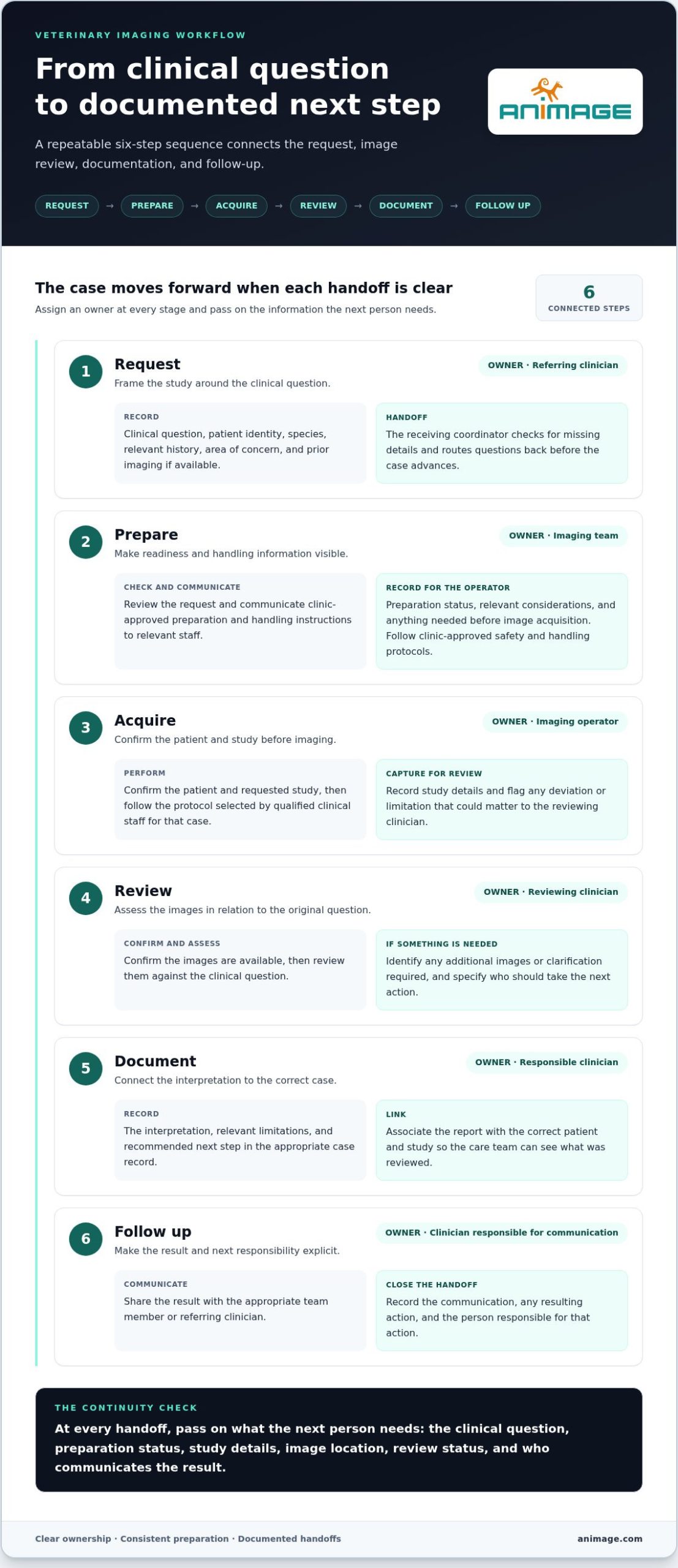
A repeatable veterinary imaging workflow gives each case a defined path while leaving clinical decisions with qualified staff. Request, prepare, acquire, review, document, follow up: each step should pass the information and responsibility needed for the next.
- Request. The referring clinician records the clinical question, patient identity, species, relevant history, area of concern, and any prior imaging that may inform the case. The receiving coordinator checks for missing details and routes questions back to the requester before the case advances.
- Prepare. The imaging team reviews the request and communicates clinic-approved preparation and handling instructions to the relevant staff. Record the patient’s preparation status, relevant considerations, and anything the operator needs before acquisition. Follow the clinic’s approved safety and handling protocols.
- Acquire. The imaging operator confirms the patient and requested study, then follows the protocol selected by qualified clinical staff for that case. Record the study details and flag any deviation or limitation that could matter during review. Protocols should reflect the case and clinical judgment, not a universal template.
- Review. The designated reviewing clinician confirms the images are available and assesses them in relation to the original question. If the study needs additional images or clarification, the reviewer identifies what is required and who should take the next action.
- Document. The responsible clinician records the interpretation, relevant limitations, and recommended next step in the appropriate case record. Link the report to the correct patient and study so the care team can see what was reviewed.
- Follow up. The clinician responsible for communicating the result shares it with the appropriate team member or referring clinician. Record the communication and any resulting action, such as further assessment or additional imaging, along with the person responsible.
Prepare the case and patient before image acquisition
Preparation is a handoff, not an assumption. The coordinator should make the clinical question, patient details, relevant history, requested study, and preparation status visible to the staff involved. Communicate instructions through the clinic’s established process, then record whether they were completed or whether an issue needs to be resolved before acquisition. Patient handling and safety remain governed by clinic-approved protocols.
Close the loop after image acquisition
Acquisition, image review, report documentation, and result communication are separate tasks. Mark each as complete in the case record rather than treating image capture as the end of the process. For cases involving more than one modality, define how the studies relate and where each result should be reviewed. This is particularly relevant when coordinating veterinary CT and X-ray systems. The American College of Veterinary Radiology Journal provides a professional reference point for developments in veterinary imaging practice.
Equipment should fit the sequence your team has defined. FIDEX+ combines cone-beam CT, digital radiography, and fluoroscopy in one compact veterinary platform. Explore the FIDEX+ multi-modality imaging platform as an equipment option to assess alongside your team’s roles and case processes.
Where veterinary imaging workflows commonly break down
A scanner can acquire images, but it cannot resolve an incomplete request, assign a staff member to prepare the patient, or ensure a report reaches the person responsible for the next clinical action. Workflow reliability depends on defined handoffs as well as equipment. A process gap can interrupt a veterinary imaging workflow even when the imaging system is functioning as intended.
A missing detail may require clarification; it does not automatically mean a clinical error occurred. A study may also have limitations that reflect the case or images acquired rather than a failure in the clinic’s process. Identify what happened, document it accurately, and avoid treating every delay or limitation as preventable.
Spot gaps in requests, preparation, and ownership
A request that identifies a body region but omits the clinical question may leave the imaging team unsure what information the referring clinician needs. The team may need to pause and request context before scheduling or preparation can proceed. Treat that clarification as a workflow checkpoint, not a reason to guess at the intended study.
Ownership gaps can be less visible but just as disruptive. If no role is assigned to communicate preparation instructions, staff may assume someone else has handled it. If the person coordinating the case is not responsible for recording completion, preparation status can remain unclear at handoff. Similar gaps occur when images are ready but no one is assigned to route them for review or notify the referring clinician.
Document responsibilities in the clinic’s workflow materials. Name the role responsible for checking request completeness, coordinating preparation, confirming the study is associated with the correct case, and communicating review status. Roles vary by staffing model. The essential point is that each task has an owner, even when one person performs several tasks.
Separate image-quality checks from workflow checks
Technical review and process review answer different questions. Technical review asks whether the images are usable for the intended clinical assessment. Process review asks whether the right case information, people, and records moved through the agreed sequence. A satisfactory process does not guarantee diagnostic image quality, and a technically adequate image set does not prove that reporting and follow-up were completed.
For technical review, qualified staff can assess case-appropriate factors such as positioning and adherence to the selected protocol. Protocol decisions should remain with qualified clinical staff and reflect the patient and clinical question. A general checklist can prompt review, but it should not replace clinical judgment or prescribe one protocol for every patient and modality.
For process review, check whether the request contained enough context, preparation instructions were communicated and recorded, images were associated with the correct case, and the report and next action were documented. Keep these checks distinct in team discussions. If image quality needs attention, review the technical factors. If a handoff failed, clarify task ownership or documentation. This separates operational gaps from clinical limitations without overstating what a revised workflow can prevent.
How to make a veterinary imaging workflow repeatable
A repeatable veterinary imaging workflow does not require a large manual. It needs a compact set of checkpoints suited to the clinic’s case mix, staffing, and available modalities. Keep the process practical during routine work: staff should be able to identify the owner, see what is complete, and know what must happen next.
Create a checklist staff can use during a case
Build the checklist around handoffs, not equipment menus. Record the case purpose, assigned roles, preparation status, image location or transfer status, review completion, and follow-up owner. Keep clinical protocols approved by the practice and appropriate to the modality and case. The checklist tracks whether the agreed process was followed; it does not replace clinical judgment or prescribe acquisition settings.
- Case: patient identifier, clinical question, requested study, and relevant context.
- Ownership: person or role responsible for coordination, acquisition, review, and communication.
- Preparation: instructions communicated and status recorded.
- Images and review: study associated with the correct case, routed for review, and marked reviewed.
- Closure: report documented, result communicated, and next action assigned or recorded as not required.
Use fields that match existing records where possible. A short checklist embedded in a familiar routine is more practical than a separate form staff must remember to find.
Make handoffs visible
A handoff is complete when the next person can act without guessing. Compare the difference:
| Undefined handoff | Documented handoff |
|---|---|
| “The images are ready.” | Study location, case identifier, and reviewer are recorded. |
| Preparation is assumed complete. | Preparation status and any unresolved issue are noted. |
| The report is filed with no communication owner. | Report status, intended recipient, and follow-up responsibility are specified. |
The details differ by practice. A clinic with a small team may assign several steps to one person; a referral hospital may distribute them across roles. Either way, each handoff needs an owner and a visible completion point.
Review workflow performance without overcomplicating it
Look for recurring process gaps, such as requests that often need clarification, missing preparation records, or reports without a documented communication step. These are indicators to investigate, not automatic proof of poor performance. Delays can have different causes from case to case, so avoid imposing benchmarks without a basis in the clinic’s workload and needs.
Ask the staff who perform the handoffs where instructions or records become unclear. Make one practical adjustment, then review whether the updated step is understandable and usable. Keep the process proportionate to the clinic’s size and case mix, and revisit it when staffing or modality use changes.
When evaluating equipment within a defined process, explore FIDEX+ multi-modality imaging. The compact veterinary platform combines cone-beam CT, digital radiography, and fluoroscopy.
Where an integrated imaging platform fits in the veterinary workflow
Equipment should support a defined clinical sequence, not stand in for one. A multi-modality platform brings different imaging options into the clinic’s equipment environment, but it does not decide which modality suits a case, assign staff responsibilities, or replace clinical protocols. Those decisions remain with the veterinary team.
Fit depends on the practice. Case mix, staffing, and existing processes shape how modalities are accessed and how images move to review. Assess equipment against those operating conditions rather than assuming that combining modalities will automatically change case flow.
Consider what multi-modality access changes operationally
Start by mapping where modality decisions happen in the existing case sequence. Which role coordinates access? Who confirms the intended study? How are the resulting images associated with the case and made available for review? Answering these questions helps a team assess whether a multi-modality system fits its routines without assuming the equipment alone resolves handoffs.
For example, a practice can consider how cone-beam CT, digital radiography, and fluoroscopy would fit its case routing and staff responsibilities. The choice of modality and protocol still depends on the clinical question and qualified clinical judgment. Access to more than one modality does not remove the need for appropriate preparation, documentation, or review.
Explore FIDEX+ as one workflow option
FIDEX+ is a compact veterinary imaging platform that combines cone-beam CT, digital radiography, and fluoroscopy. It is designed for veterinary clinics, referral hospitals, and research institutions, without requiring dedicated CT-room construction. These are equipment characteristics, not a promise of a particular workflow result. How the platform fits depends on the practice’s cases, staff roles, and established procedures.
Teams can assess its place in their workflow by considering how the three modalities relate to their imaging needs and how each study would move through request, acquisition, review, and follow-up. FIDEX+ provides imaging equipment; case-specific clinical decisions remain with veterinary professionals.
For a closer look at the platform’s modality combination and intended settings, explore Animage’s veterinary imaging platform.
Put the next workflow improvement into practice
A useful next step is to review one recent imaging case with the people who handled it. Identify where someone had to infer what to do, where information was difficult to find, or where the next action was unclear. Choose one point to make explicit in the clinic’s process, then use team feedback to see whether the change fits daily work.
Build the veterinary imaging workflow around the people, case types, and resources your practice actually has. A process staff can follow consistently is more useful than a complex procedure that exists only on paper. Consider equipment against that defined process rather than treating it as a substitute for clear responsibilities.
To evaluate equipment fit, explore Animage’s FIDEX+ veterinary imaging platform and consider how its cone-beam CT, digital radiography, and fluoroscopy capabilities align with your clinic’s imaging needs. Involve the team that will use the system, map the handoffs, and improve one step at a time.
Frequently Asked Questions
How long does a veterinary imaging workflow take?
There is no fixed duration for a veterinary imaging workflow. Timing depends on the case, modality, patient preparation, staff availability, and how images are reviewed. A straightforward study may move through the clinic’s stages differently from a case requiring additional preparation or external interpretation. Track where time is spent, such as waiting for missing information or a review decision, and use those observations to set realistic expectations.
Can one imaging workflow cover different veterinary modalities?
Yes. A clinic can use one overarching process for requests, case identification, image handling, reporting, and follow-up, while keeping acquisition procedures specific to each modality. For example, a radiography study and a CT examination can share a case-tracking approach but require different clinical protocols and preparation. Make modality-specific instructions easy to find within the shared process, so a general checklist does not blur important differences or replace clinical judgment.
What information should an imaging request include?
An imaging request should identify the patient, describe the clinical question, and specify the requested study or area of concern. Include relevant history, laterality when applicable, previous studies if available, and the referring clinician’s contact details for clarification. For instance, noting which limb is affected gives the team useful context beyond “lameness.” Include urgency or scheduling constraints when relevant, but leave modality and protocol decisions to qualified clinical staff.
What is DICOM in a veterinary imaging workflow?
DICOM, or Digital Imaging and Communications in Medicine, is a standard for storing and exchanging medical images and associated information. In veterinary imaging, it can support movement of studies between compatible modalities and image-management systems. DICOM compatibility alone does not ensure that every system will exchange or display every item as intended. Understand how your systems handle image transfer and case identification, and define a process for addressing transfer problems.
How can a clinic reduce missed handoffs in imaging cases?
Assign each handoff to a role and make completion visible in the clinic’s usual record or tracking method. A practical safeguard is a closed-loop acknowledgement: the person receiving a study or report marks it received, or flags that something is missing. For example, do not treat “sent for review” as equivalent to “review received.” Periodically compare the process record with staff experience to find steps that are easy to overlook during busy periods.
What happens if images are incomplete or difficult to review?
The reviewing clinician assesses whether the available images are adequate for the clinical question and determines the appropriate next step. That may involve requesting additional images, noting a limitation in the report, or considering other relevant information. The team should communicate the issue to the responsible clinician and document the decision. Do not assume that every difficult-to-review study requires repeating acquisition; the response depends on the case and clinical judgment.
Does improving an imaging workflow require new equipment?
No. A clinic can often begin by clarifying who checks requests, how case status is recorded, and how review or follow-up is acknowledged. New equipment may be worth considering if existing capabilities do not fit the practice’s imaging needs, but it is not a substitute for defined responsibilities. First identify the operational problem, then decide whether it relates to process, equipment, or both. This keeps purchasing decisions tied to actual clinical and workflow requirements.