Advanced imaging supports revenue when it improves clinical decisions and strengthens the economics of a case. A sound approach to increasing veterinary clinic revenue with advanced imaging rests on three factors: keeping suitable cases in-house, helping owners understand recommended care, and providing a pathway to treatment after diagnosis. CT and MRI answer different diagnostic questions, but their financial contribution depends on appropriate case volume and a workflow that fits the practice.
The investment question is practical: can imaging activity and related care cover the equipment’s total operating costs over time? There is no universal break-even point. Case mix, referral demand, staffing, scheduling and the services connected to a diagnosis all affect the calculation.
This article explains how MRI and CT support diagnosis, client acceptance and referral relationships, then shows how to assess the revenue opportunity and the conditions that shape break-even.
Key Takeaways
- Match MRI or CT to the diagnostic question, then consider how the findings could shape treatment planning.
- Assess technology adoption against your clinic’s case mix, referral demand, staffing and workflow.
- Estimate break-even by weighing expected imaging activity and follow-on care against the full costs of operating the system.
- Use clear imaging findings to help pet owners understand recommended care, and consider whether FIDEX+ fits your clinic’s imaging goals.
How MRI and CT support diagnosis and treatment planning
Advanced imaging can generate revenue when it answers a clinical question and informs the next step. CT provides cross-sectional views that can help assess complex anatomy and trauma. MRI can show soft-tissue detail, including causes of persistent neck pain in dogs. Veterinarians use imaging findings to localise disease, refine the diagnostic plan and discuss treatment options with owners. A diagnosis may also lead to further care within the practice.
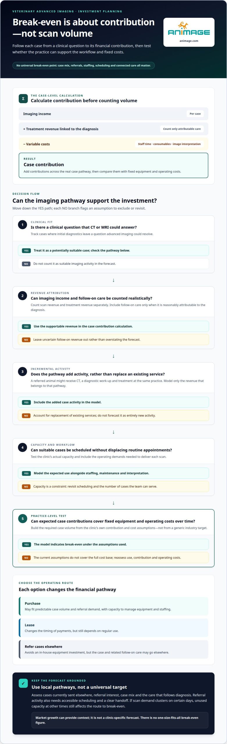
Buying equipment, leasing and referring cases elsewhere suit different operating conditions. Purchasing may fit a clinic with predictable case volume, referral demand and the capacity to manage equipment and staffing. Leasing changes the timing of payments, but still depends on regular use. Referring cases avoids an in-house equipment investment, but the case and related follow-on care may go elsewhere. Compare the options using expected eligible cases, operating costs and the value of treatment retained.
For Polish clinics, radiation-safety requirements depend on the imaging modality. CT and digital radiography use ionising radiation, so installation and operation must account for applicable Polish radiation-protection requirements. MRI does not use ionising radiation and has its own safety procedures. Identify the relevant modality first, then factor the requirements for the specific system and clinical workflow into planning.
Poland-wide demand is shaped by local case mix and referral patterns, not by one uniform figure. A clinic can assess its opportunity by tracking cases where initial diagnostics leave a question that CT or MRI could answer, then noting referrals sent elsewhere. The research reports that 45% of veterinary practitioners prefer CT for complex diagnostics. That figure indicates interest in CT for complex cases, not a measure of demand in Poland.
Pet insurance can affect an owner’s ability to consider diagnostic imaging, but figures from the United States do not describe the Polish market. Revenue growth therefore depends on local case acceptance, appropriate scan volume and care following diagnosis. Assess each case by its diagnostic purpose, referral source and subsequent care. Imaging is financially relevant when the clinical pathway generates enough activity to support the investment and operating model.
Private veterinary care and technology adoption
Adopting imaging technology in private veterinary care depends on clinical need and how the practice manages each visit. Digital radiography can answer common first-line questions; CT or MRI may be appropriate when two-dimensional images leave uncertainty. For example, a complex trauma case may need cross-sectional imaging to clarify anatomy before treatment planning. The opportunity is not simply to add equipment, but to connect the capability with appropriate cases, referral pathways and follow-on care.
Cost is a central consideration. An imaging system involves more than the acquisition decision: the clinic must account for staff time, maintenance, room requirements, workflow disruption and ongoing operation. Adoption may be supported by cases currently referred elsewhere, requests from other practices and the ability to schedule scans without disrupting routine visits. A clear case pathway helps: identify the clinical question, decide whether imaging can answer it, and plan how the results will inform treatment. This grounds the investment decision in veterinary practice rather than technology alone.
For veterinary CT, MRI and ultrasound in Poland, scan prices depend on the case and are provided after a quote request. Modality, patient size, sedation or anaesthesia, and image interpretation all affect the estimate. These factors also shape how clinics explain the diagnostic value and likely next steps to pet owners.
Staffing and operating costs vary by modality and workflow. Account for trained operators, veterinarian time for case selection and interpretation, and any external image-reading support. Technicians may operate imaging systems using suitable software, allowing veterinarians to focus on other responsibilities. The operating model must also cover scheduling, patient preparation, image transfer, equipment upkeep and staff training. Each factor affects capacity, not just the scan itself.
To assess adoption, map visits that currently lead to external imaging, identify referral interest from other practices, and estimate how in-house access could change case flow. Then compare expected use with staffing and operating demands. Advanced imaging can expand diagnostic capability, but works best when clinical demand, workflow and investment assumptions align.
Break-even cases and profitability
Break-even depends on contribution, not scan volume alone. For each case, calculate imaging income and treatment revenue linked to the diagnosis, then subtract variable costs such as staff time, consumables and image interpretation. Compare the result with fixed equipment and operating costs. The required case volume should come from this calculation, not a generic industry target.
Build the model around actual clinic pathways. For example, a referred animal arrives for CT, receives a diagnostic work-up, then proceeds to treatment at the same practice. Count the scan and follow-on care separately, including only revenue reasonably attributable to that pathway. Account for cases that replace existing services instead of adding activity. Otherwise, the forecast may overstate the financial gain.
Next, test capacity. Estimate how many suitable cases the team can schedule without displacing routine appointments, and include staffing, maintenance and interpretation in the operating model. A specialty service may attract referrals from other practices, but referral activity requires accessible scheduling and a clear handoff. If scan demand clusters on certain days, unused capacity at other times still affects the path to break-even.
Market growth offers context, not a clinic forecast. Veterinary diagnostic imaging grew by 4.6% in iVET360’s 2026 benchmark report. That growing category does not establish local demand or prove that a particular offering will be profitable. In Poland, clinic-level case mix, referral relationships, owner acceptance and available capacity determine the relevant revenue assumptions.
Use sensitivity analysis before committing: model lower, expected and higher case volumes, then recalculate contribution after operating costs. Identify which assumption has the greatest effect, such as referral conversion or follow-on treatment. Keep equipment costs in PLN and separate the one-time investment from recurring expenditure. This makes the break-even point visible and shows whether advanced imaging can support animal health and clinic profitability under realistic operating conditions.
Client understanding and treatment acceptance
Imaging gives veterinarians visual evidence to connect clinical findings with a suspected disease. That evidence can help owners understand why treatment is recommended, beyond a verbal explanation alone. A scan does not make the decision for them. It gives the veterinarian a clearer basis for discussing what the images show, what remains uncertain and how each treatment option relates to the patient’s condition.
Consider the documented MRI case of a cat with a mass affecting the left external ear canal. The images showed extension towards the middle ear and surrounding tissues, while the middle and inner ear structures remained preserved. Surgery later confirmed squamous cell carcinoma with secondary infection. The case illustrates how imaging can define the extent of disease and support a focused discussion of treatment planning.
For a useful consultation, present the images alongside the clinical question. Explain the relevant anatomy in plain language, then connect the finding to the proposed next step. For example, show where a mass extends before outlining how surgery may address it. This approach builds understanding without suggesting that an image alone determines the right treatment or outcome.
Veterinary practices can make discussions more consistent by documenting the owner’s questions, the finding explained and the agreed plan. This record helps the clinical team maintain continuity between appointments. It also helps staff distinguish uncertainty about the diagnosis from concerns about treatment, which require different explanations.
As diagnostic options expand, the ability to explain findings becomes part of the imaging workflow. Clear images can support owner understanding, but acceptance still depends on the case, the proposed care and the owner’s decision. Clinics can strengthen the conversation by pairing each image with a specific explanation of its clinical relevance. To assess how imaging fits that workflow, review veterinary imaging options for your practice.
Animage
Animage develops and sells FIDEX+, an imaging platform for veterinary clinics considering in-house diagnostic capability. It combines cone-beam CT, digital radiography and fluoroscopy in one compact device. The multi-modality system brings several imaging functions together, helping a practice assess how advanced imaging could fit its clinical workflow and investment plan.
FIDEX+ is purpose-built for animals and serves veterinary clinics, referral hospitals and research institutions. It may suit a practice evaluating CT alongside radiography and fluoroscopy, rather than planning around a single imaging modality. Consider the cases your team aims to assess, the workflow they require and how imaging could support treatment planning.
For a practical discussion of FIDEX+ and how it could fit your clinic’s imaging plans, contact Animage.
Make the Next Decision Practical
Before expanding imaging capacity, turn the financial model into an operational decision. Identify the cases your clinic aims to serve, how imaging fits existing appointments and who will coordinate each stage. This gives your team a shared basis for assessing whether the proposed workflow is manageable alongside current veterinary care.
A disciplined approach to increasing veterinary clinic revenue with advanced imaging treats revenue as the result of appropriate case selection and consistent delivery, not a guaranteed outcome of owning equipment. Review the assumptions with clinical and operational leaders, then decide what evidence would support moving forward. Clear criteria keep the decision grounded in your clinic’s priorities.
For a discussion about how imaging equipment could fit your practice’s plans, contact Animage to discuss imaging. A focused conversation can help you consider the next step with clarity and confidence.
Frequently Asked Questions
How can advanced imaging increase revenue at a veterinary clinic?
Advanced imaging can contribute revenue through diagnostic scans, referrals and care following diagnosis. For example, a clinic may receive a complex case from another practice, perform appropriate imaging, then provide related treatment. The financial effect depends on the services the clinic delivers and how it manages the case. A strategy for increasing veterinary clinic revenue with advanced imaging should track both scan activity and subsequent care.
What is the return on investment and payback period for veterinary imaging equipment?
Calculate return by comparing total equipment ownership and operating costs with the contribution from imaging and related care. The payback period depends on case volume, referral flow, staff capacity, maintenance and the proportion of cases that proceed to treatment. Model different levels of use rather than relying on a single forecast. Separate new cases from services that would have occurred anyway, so projected returns reflect added activity.
How can a veterinary clinic market advanced imaging and increase referrals?
Build referrals through clear clinical communication with other practices. Explain which cases the imaging capability can support, how referring veterinarians receive findings and how the clinic coordinates the next stage of care. Share relevant case examples with appropriate permission, and keep referral updates timely and concise. A consistent process gives practices a practical reason to refer suitable cases and helps owners understand why imaging is recommended.
What are the risks and hidden costs of adding advanced imaging services?
The main risks are underused capacity, workflow disruption and operating costs that exceed the contribution from scans. Account for staff training, preparation time, maintenance, interpretation, room requirements and the effect of scheduling on other appointments. For CT and digital radiography, include radiation-safety processes in operational planning. A capacity model should also consider downtime and cases requiring external expertise, then test whether the service remains workable when demand is lower.