Does every difficult imaging case call for CT, or can a well-positioned radiograph answer the question? Choosing a veterinary CT and x-ray system starts with the diagnostic task, not the appeal of advanced technology. Digital radiography gives clinicians a fast view of structures such as bones and the size or position of organs. CT adds cross-sectional detail when overlapping anatomy makes a case harder to assess.
It’s useful to weigh that extra detail against room layout, staffing and daily workflow. CT and radiography answer different questions, so they aren’t competing versions of the same tool. A clinic may use radiography for an initial examination, then consider CT when further detail is needed. Whether the equipment is separate or integrated depends on the clinic’s case mix and how patients move through the imaging process.
This article explains what each modality shows, when CT may add useful information and how to assess a combined setup. It also introduces FIDEX+, which brings veterinary cone-beam CT, digital radiography and fluoroscopy together in one device without requiring dedicated CT-room construction. Use the practical criteria below to match imaging capability to clinical tasks and clinic workflow.
Key Takeaways
- Match each imaging method to the diagnostic question: radiography produces projection images, while CT provides cross-sectional image data.
- Compare image format, clinical question and workflow role to clarify when each modality may contribute.
- Assess an integrated veterinary CT and x-ray system against your case mix, room layout, staffing and existing equipment.
- Plan imaging around clinical requirements first, then consider equipment preferences and facility constraints.
- FIDEX+ combines veterinary cone-beam CT, digital radiography and fluoroscopy for clinics, referral hospitals and research institutions.
What Is a Veterinary CT and X-Ray System?
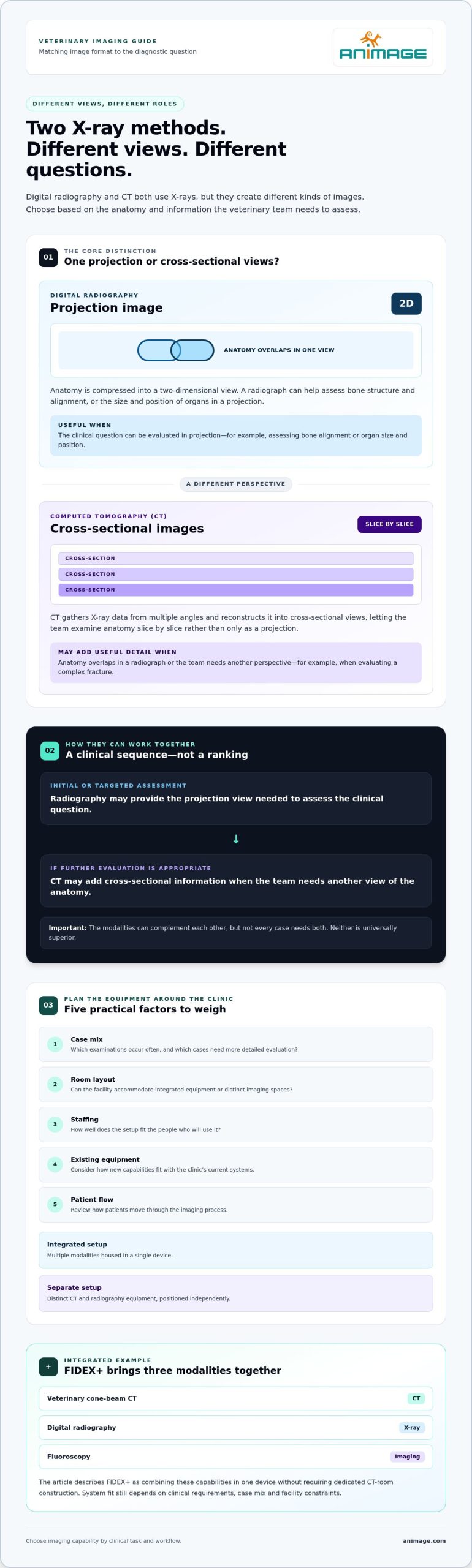
A veterinary CT and x-ray system provides equipment for two imaging methods that use X-rays but produce different kinds of images. Digital radiography creates projection images, where anatomy is compressed into a two-dimensional view. CT acquires X-ray data from multiple angles and reconstructs it into cross-sectional images. The distinction matters because clinicians choose an imaging method to answer a particular diagnostic question, not simply to obtain more advanced equipment.
What does “CT and X-ray system” mean in veterinary imaging?
The phrase can describe a clinic’s overall imaging capability, not necessarily one machine. An integrated platform houses multiple modalities in a single device. A separate setup uses distinct CT and radiography equipment, each with its own place in the facility and workflow. In either arrangement, CT builds cross-sectional image data, while digital radiography produces projection images for veterinary assessment.
The image formats support different ways of examining anatomy. A radiograph can help assess bone structure, alignment, or the size and position of organs in a projection. CT separates anatomy into cross-sections, which can help evaluate complex fractures or structures that are difficult to distinguish when they overlap in a projection. That doesn’t make radiography inadequate or CT the default. Each method has a defined role.
Why consider more than one imaging method?
Start with the clinical question and the anatomy of interest. A projection image may provide the view needed for an initial assessment. A more complex question may call for cross-sectional detail. The appropriate choice depends on the patient and the information needed to guide the next clinical decision. Neither modality is universally superior, and not every case requires both.
Case mix shapes the practical value of additional imaging. A clinic that regularly evaluates a broad range of cases may have different imaging requirements from a practice whose routine needs are met by radiography. Review which examinations occur often, which cases need more detailed evaluation and how patients move between imaging steps. Those patterns help distinguish a clinical requirement from a preference for a particular system.
The equipment arrangement matters too. An integrated platform brings modalities together in one device; separate systems let a clinic position CT and radiography equipment independently. Space, existing equipment, staffing and patient flow all affect how well either arrangement fits. Integration brings capabilities together, but the setup still needs to work for the people using it and the cases they see.
Use these practical decisions to plan imaging capability: identify what each modality contributes, consider how integrated or separate equipment fits the clinic workflow, and assess the facility requirements. This is not a technical comparison of every scanner specification. The aim is to connect diagnostic tasks with a workable system plan, so equipment choices follow clinical needs.
How Veterinary CT and Digital X-Ray Serve Different Diagnostic Tasks
A veterinary CT and x-ray system supports distinct diagnostic tasks, even though both modalities use X-rays. Digital radiography records anatomy in a projection image. CT gathers data from multiple angles and reconstructs cross-sectional views. The choice isn’t a simple ranking from basic to advanced. It depends on what the veterinary team needs to assess and how much anatomical detail the question requires.
Image format: Digital radiography produces projection images; CT produces cross-sectional views reconstructed from X-ray data.
Typical clinical question: Radiography can show bone alignment or organ size and position in a projection. CT provides another perspective when the team needs to examine anatomy across slices rather than in a single projection.
Workflow role: Radiographs may support an initial or targeted assessment. CT can add information when further evaluation is appropriate. The modalities may complement each other, but not every case calls for both.
What questions can digital radiography help address?
Radiography gives the veterinary team a projection-based view of the area being examined. Images may help assess a bone’s alignment or the relative size and position of structures in the chest or abdomen. This view can be useful when the clinical question can be evaluated in projection. Overlapping anatomy is part of how the image is formed, not a reason to dismiss the method.
Positioning and the selected views affect what anatomy is visible, so consider radiography in the context of the examination and the question being asked. For a closer look at its role in practice, see this guide to veterinary digital radiography.
What additional perspective does veterinary CT provide?
CT organizes data into cross-sectional images, allowing the team to examine anatomy slice by slice rather than only as a projection. This can provide a different view of structures that overlap in a radiograph. It doesn’t make every finding clearer or make radiography unnecessary. CT is a complementary option when the attending team determines that cross-sectional information is relevant to the case.
For example, a projection image may answer the initial question, while the team needs to assess the anatomy from another perspective. CT may add that perspective; whether it’s warranted depends on the patient, the clinical question and the information needed. The attending veterinary team determines the appropriate method for each case. Equipment planning should support that clinical judgment, not substitute for it.
For a clinic considering how multiple modalities could fit within one workflow, Animage’s veterinary imaging platform offers an integrated equipment option to evaluate.
Integrated or Separate Veterinary CT and X-Ray Equipment?
A clinic can bring CT and radiography together in one platform or operate them as separate systems. Neither arrangement is automatically more efficient. The right fit depends on the imaging the clinic performs, how staff and patients move through the facility, and what equipment is already in place. Integration changes the equipment layout; it doesn’t guarantee shorter examinations, lower costs or fewer staffing requirements.
What can an integrated imaging platform change operationally?
With multiple modalities in one device, the clinic plans around a shared equipment footprint rather than separate CT and radiography units. That can affect room use, patient transfers, scheduling and staff routines. Treat these as planning considerations, not guaranteed workflow gains. Map how examinations are prepared and performed, and where image review takes place.
FIDEX+ is an example of this configuration. It combines veterinary cone-beam CT, digital radiography and fluoroscopy in a compact platform designed for animal imaging. It does not require dedicated CT-room construction. This can inform facility planning, but the intended room, staff workflow and other operational requirements still need consideration.
Evaluate the full imaging pathway, not just the equipment footprint. Consider whether the same team will use the modalities, whether cases commonly require more than one imaging method and whether a shared setup fits the clinic’s examination schedule. Integration is useful when the configuration supports actual clinical and operational needs.
When might a clinic consider separate equipment?
Separate systems can make sense when a clinic already has radiography equipment that meets its needs, or when CT and X-ray are used in different service patterns. Independent units may give the clinic more choice about equipment placement and future expansion. Weigh that flexibility against the space and coordination required to operate separate systems.
Compare the options against the clinic’s actual conditions:
- Case mix: Identify which imaging tasks are routine and which arise less often.
- Room layout: Account for available space, patient movement and how staff access each system.
- Existing equipment: Consider whether current radiography capability remains useful in a separate setup.
- Staffing and workflow: Map who operates each system and how examinations fit into daily schedules.
- Expansion needs: Consider whether future imaging requirements may call for additional or different equipment.
CT doesn’t always require a dedicated CT room. FIDEX+ does not require dedicated CT-room construction, though each clinic still needs to plan how the equipment will fit its facility and workflow. An integrated setup may suit one practice; separate systems may better serve another. Make the decision based on clinical requirements, space and staff capacity, rather than assuming one layout is superior. Explore FIDEX+ imaging equipment as an example of integrated veterinary imaging.
How to Plan a Veterinary CT and X-Ray Workflow
Plan around the work the clinic needs to perform, then assess how imaging will fit into the facility and daily routines. A clear sequence helps separate clinical requirements from equipment preferences. It also brings practical constraints to light before a system choice is made.
- List the clinic’s diagnostic questions. Identify the imaging questions the veterinary team encounters regularly. Separate routine radiography needs from cases where cross-sectional views may add useful information. Base this review on the clinic’s actual caseload and referral patterns, not an assumed scan volume.
- Define the clinical requirement. For each imaging task, note what the team needs to assess and what information the current workflow provides. Distinguish that need from preferences such as having a particular modality in-house or combining equipment. An equipment feature is valuable when it supports a defined clinical or operational requirement.
- Map the physical workflow. Review the available room, access routes, patient movement and the positions staff need during preparation and imaging. Consider how a patient moves from examination to imaging and back, and whether equipment placement creates avoidable transfers or congestion.
- Trace staff and image-review steps. Identify who prepares the patient, operates the equipment and reviews the resulting images. Map where images are assessed and how findings enter the clinic’s existing processes. Focus on the handoffs staff must manage rather than assuming a particular software platform or connectivity feature.
- Assess the selected system in context. Room-planning needs depend on the equipment and installation context. Compare the system’s physical requirements with the facility plan, existing imaging equipment and staffing model. Revisit the workflow if a combined setup changes where examinations take place or who performs each task.
Start with diagnostic needs and case mix
Case mix should guide capability planning. A clinic whose routine imaging questions are largely addressed by radiography may prioritize that workflow differently from a referral hospital managing a broader range of cases. The point isn’t to predict a precise number of examinations. It’s to understand which tasks occur, which may require an additional imaging perspective and how often the team needs to coordinate those steps.
Review space, staffing and image workflow
Walk through a typical patient journey, from arrival in the imaging area to image review and the next clinical step. Check whether staff can move and work safely around the equipment, where the patient is positioned and how the team handles a case that needs more than one imaging method. Include existing routines, not just the proposed equipment layout. A floor plan alone won’t show every handoff.
For a veterinary CT and x-ray system, the most useful plan connects diagnostic tasks with staff movement and available space. Equipment labels don’t define the workflow. Explore FIDEX+ integrated imaging equipment as one option when assessing how modalities could fit within a clinic’s imaging plan.
Where FIDEX+ Fits in a Veterinary CT and X-Ray System Plan
Once a clinic has identified the imaging tasks it needs to support, it can consider an integrated platform alongside separate equipment. FIDEX+ is Animage’s compact veterinary imaging platform, designed for veterinary clinics, referral hospitals and research institutions. It combines cone-beam CT, digital radiography and fluoroscopy in one device. It does not require dedicated CT-room construction, which is a practical consideration for facilities assessing how advanced imaging could fit their available space.
What modalities does FIDEX+ combine?
The platform brings together three imaging approaches. Cone-beam CT reconstructs cross-sectional image data from X-rays acquired around the patient. Digital radiography creates projection images. Fluoroscopy provides X-ray imaging over time, offering a view of movement rather than a single static projection. These methods serve different purposes. Having them in one device does not mean every case requires each one or guarantees a particular diagnostic result.
For a clinic, the relevant question is how those capabilities relate to established examination pathways. Consider whether the team’s case mix calls for these types of imaging, how the modalities could fit into daily routines and whether a shared platform aligns with staff roles and the facility layout. Integration brings imaging methods together physically, but clinical decisions remain specific to each patient and case.
How can a clinic assess whether the platform fits?
Use the same planning criteria applied to any veterinary CT and x-ray system. Start with clinical requirements, then consider equipment preferences. A combined platform may be relevant when multiple modalities fit a clinic’s diagnostic needs, but no single configuration suits every practice. Compare the expected role of each modality with existing imaging capabilities and the way cases move through the facility.
- Case mix: Identify the imaging questions the team encounters and which modalities may be relevant.
- Workflow: Map patient handling, staff responsibilities, examination scheduling and image review.
- Room footprint: Consider where the platform would sit and how people and patients would move around it.
- Existing equipment: Assess how an integrated device would complement current imaging resources and processes.
FIDEX+ combines its modalities in a compact format and does not require dedicated CT-room construction. Consider this in facility planning alongside the specific site and installation context. The useful comparison is not simply integrated versus separate. It is whether the equipment configuration supports the clinic’s clinical requirements, staff workflow and available space.
If that combination aligns with your planning priorities, explore the FIDEX+ veterinary imaging platform as a practical next step in assessing integrated imaging.
Turn Imaging Priorities Into a Practical Plan
Translate diagnostic priorities into an equipment plan the clinical team can use day to day. Define the cases and workflows the system needs to support, then evaluate the room layout, staff roles and existing imaging capability against those needs. A veterinary CT and x-ray system should fit the clinic’s operating conditions, not an abstract idea of what an advanced imaging service looks like.
FIDEX+ is one integrated option to assess. It combines cone-beam CT, digital radiography and fluoroscopy in a compact platform that doesn’t require dedicated CT-room construction. Consider how that configuration relates to your facility and imaging plans, without assuming one setup is right for every practice.
Explore the FIDEX+ veterinary imaging platform to see how its combined modalities may fit your clinic’s diagnostic workflow. Start with the questions your team needs imaging to answer, then use them to shape a practical equipment plan.
Frequently Asked Questions
Is veterinary CT the same as a standard X-ray?
No. Veterinary CT uses X-rays to create cross-sectional image data, while conventional radiography produces projection images. They use related technology but show anatomy differently. The team may consider whether a projection can address the question or whether views across multiple sections could add relevant information. The animal’s anatomy, examination purpose and clinical context all matter. A veterinary professional determines which imaging method is appropriate for the individual case.
Can one veterinary system include both CT and X-ray?
Yes. Some platforms combine CT and digital radiography, while other clinics use separate equipment. FIDEX+ is Animage’s veterinary imaging platform, combining cone-beam CT, digital radiography and fluoroscopy in one device. For a clinic evaluating a veterinary CT and x-ray system, consider how that arrangement fits its examination pathways, available space and existing equipment. A combined platform is one configuration, not a requirement for every practice.
Does a veterinary CT system always need a dedicated CT room?
No. Room requirements vary with the selected system and installation context. FIDEX+ is a compact platform that does not require dedicated CT-room construction, but the clinic still needs to plan where the equipment will sit and how staff and animals will move around it. A room review should consider access, working space and the clinic’s established imaging process. Don’t assume another CT system will have identical facility requirements.
Does every animal need sedation or anaesthesia for veterinary CT?
No universal rule applies. The decision depends on the animal, the examination and the veterinary team’s clinical judgment. Equipment alone doesn’t determine how a patient should be managed. The team considers the animal’s individual needs and what the imaging examination requires. This FAQ can’t determine what is appropriate for a particular patient. The attending veterinary professionals make that decision for each case.
Is veterinary CT appropriate for every diagnostic question?
No. Imaging methods provide different information, and a more advanced system isn’t automatically the right choice for every question. The veterinary team considers the case, the anatomy being assessed and the information needed to guide the next step. If the clinical question can be addressed through an existing examination pathway, additional imaging may not be indicated. Equipment capability supports clinical work, but it doesn’t replace professional judgment.
What information should accompany a veterinary imaging referral?
A useful referral gives the receiving veterinary team context for the imaging request. This may include the clinical question, relevant history, examination findings and any prior images that help explain the reason for referral. Noting which area or concern needs evaluation can clarify the purpose of the study. Referral procedures vary between clinics, so use the established process for sharing the information and images relevant to the case.
How should a clinic think about radiation safety when using veterinary CT or X-ray?
Radiation safety relies on appropriate equipment use, trained personnel and protocols applicable to the clinic and system. In practical planning, define who performs each imaging task and how staff follow the facility’s procedures during examinations. Don’t assume that a workflow suitable for one modality or system automatically applies to another. Veterinary teams should follow the requirements and safety procedures relevant to their equipment and facility, without relying on general guidance as a substitute for those protocols.