Case Study I: Diagnose
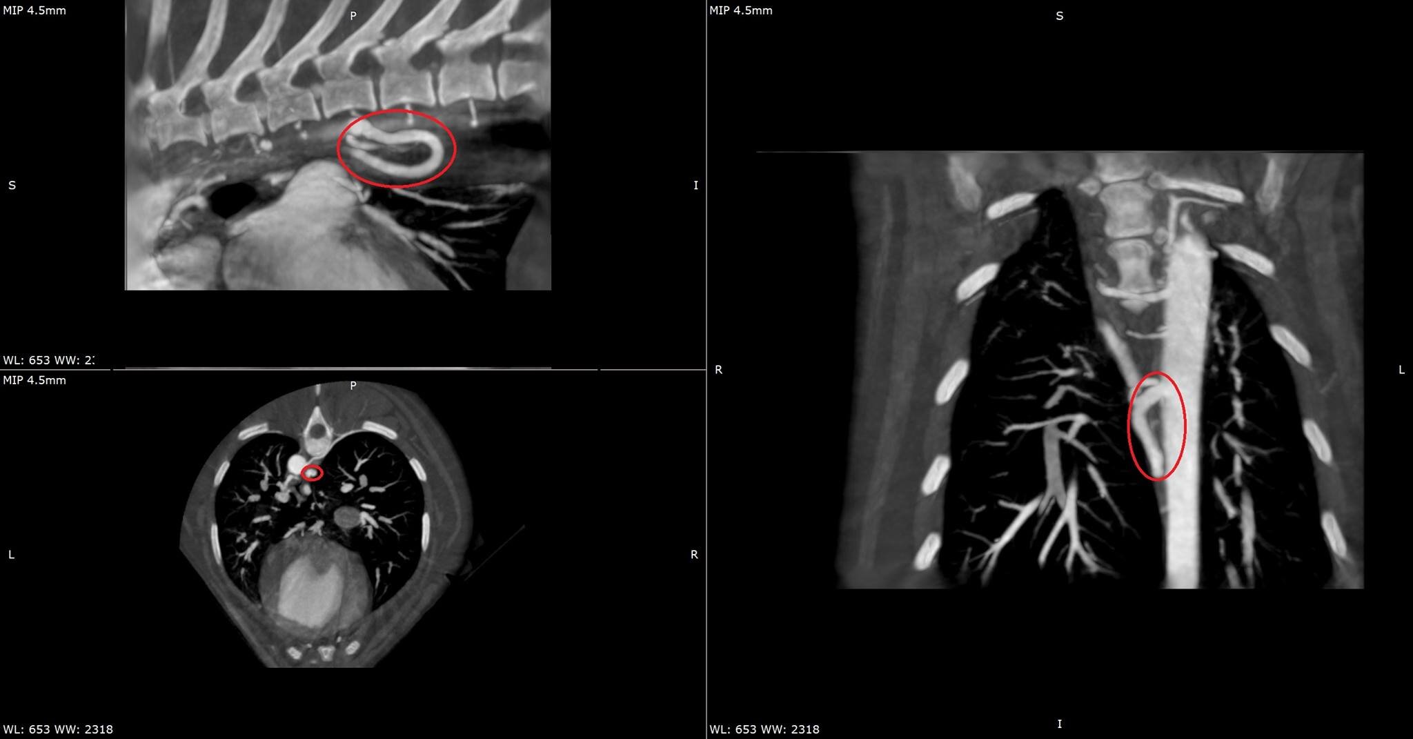
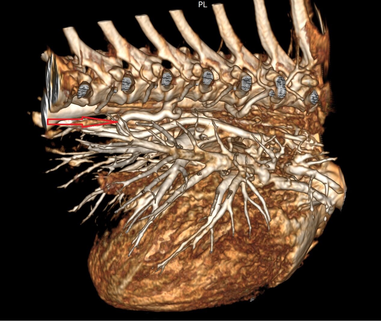
Cavalier with suspected PDA. Angiography has been carried out to identify a case with more than a typical PDA. Additional imaging including angio-CT has been carried out. The torturous vessel has been marked with a red arrow for reference.

Case Study II: Surgery Planning
A 2-year old German Shepherd with a need for a bilateral corrective osteotomy of the radius. Surgery can be planned based on CT images above. Post-surgery scans can be seen on the right,
